Download
1 / 17
170 likes | 317 Vues
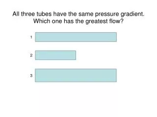
All three tubes have the same pressure gradient. Which one has the greatest flow?. 1. 2. 3. Test # 2 Monday covers. Nervous system Electrophysiology Anatomy and pathways Sensory physiology Visual System Auditory and Vestibular System Muscle Physiology

E N D
All three tubes have the same pressure gradient. Which one has the greatest flow? 1 2 3
Test # 2 Monday covers • Nervous system • Electrophysiology • Anatomy and pathways • Sensory physiology • Visual System • Auditory and Vestibular System • Muscle Physiology • Skeletal muscle and smooth muscle • Control of Movement
1QQ 8:30 Nov 2Write each letter and circle the letter of correct statements. • When blood leaves a capillary in the anterior pituitary, the next vessel it enters is a systemic venule. • A short large diameter tube would have a greater resistance than a long small diameter tube. • If one doubles the viscosity of a fluid, a pressure gradient twice as large would be necessary to maintain the same flow. • If one doubles the radius of a tube and kept the pressure gradient constant, flow through that tube would double. • Arterioles and capillaries are classified as resistance vessels.
1QQ #23 for 10:30Write each letter and circle the letter of correct statements. • When blood leaves a capillary in the anterior pituitary, the next vessel it enters is a systemic venule. • A short large diameter tube would have a lower resistance than a long small diameter tube. • The two most important factors that influence blood flow are the length of the vessel and the viscosity of the blood. • If one doubles the radius of a tube and kept the pressure gradient constant, flow through that tube would double. • Arterioles and capillaries are classified as exchange vessels.
1QQ #23 for 11:30Write each letter and circle the letter of correct statements. • When blood leaves a capillary in the anterior pituitary, the next vessel it enters is a portal vessel. • A short large diameter tube would have a greater resistance than a long small diameter tube. • The two most important factors that influence blood flow are pressure gradients and viscosity. • If one doubles the radius of a tube and kept the pressure gradient constant, flow through that tube would quadruple. • Arterioles are classified as resistance vessels.
S 8 F=Q=ΔP/R Flow = Pressure gradient/Resistance from Ohm’s Law (V=IR) R = 8Lη/πr4 R = 8Lη/πr4 Q=ΔP πr48LηPoiseulle’s equation Double radius … 16x flow Half radius….1/16th flow
S 8 Radius of arterioles regulates Q to organs Smooth muscles determine radius
Formed elements Blood volume = 5 liters = plasma + Formed Elements S 3 Figure 12.01 Serum = plasma – clotting factors Entering and Exiting the blood Discontinuous capillaries in bone marrow, spleen, & liver permit erythrocytes to enter and exit blood. Components…… Hct = percentage of blood volume occupied by RBCs. Hct regulated by Erythropoietin (EPO) from kidney. Anemia
S 1 Cardiac Output = Heart Rate X Stroke VolumeWhat regulatesheart rate?CO = HR x SV5L/min = 72 beat/min x 70 ml/beat What regulatesStroke Volume? The Cardiac Cycle animation
Bicuspid=Mitral Tricuspid“RST” Figure 12.07 Heart sounds produced by valve closings S 4 Four valves, only two heart sounds: lub dub What causes SL valves to close? SemilunarValves Animation
Atrioventricular valves and chordae tendonae Which is L and R ventricle?
S 4 Heart sounds produced by valve closings Problems with valves: ….Stenosis (narrowing) →Heart Murmurs (turbulent flow past a constriction) note: origin of neonatal heat murmurs (foramen ovale) ….Prolapse (eversion) allows backflow (also generates murmurs) Heart murmurs ≠ heart sounds Animation
Cardiac myofibers Action potentials spread from cell to cell at intercalated discs
S 3 Figure 12.13 Plateau phase Cardiac Myofiberaction potential Long refractory period prevents summation in cardiac myofibers CardiacMyofiber Cardiac myofibers are not spontaneously active so where does the heartbeat originate?
Heartbeat begins with an action potential in the Sino-Atrial Node SA node cells do not have stable resting membrane potential, spontaneously depolarize and produce an AP, are Pacemaker cells
S 5 Figure 12.14 Cardiac Pacemakeraction potential Ectopic Pacemaker Locations other than SA Node Pacemaker Cells in Conducting System: SA Node andBundle of His These cells set the rhythm & control Heart Rate.
S 4 Figure 12.11 S 5 SA node cells do not have stable resting membrane potential, spontaneously produce AP, are Pacemaker cells