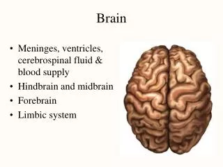
brain
brain. From the brain and from the brain only, arise our pleasures, joys, laughter and jests, as well as our sorrows, pains, grief's and tears. ~Hippocrates. Marilyn Rose. meninges. 3 membranes Outer - Dura mater- folds house the following: flax cerebri- cerebral hemisphere

brain
E N D
Presentation Transcript
brain From the brain and from the brain only, arise our pleasures, joys, laughter and jests, as well as our sorrows, pains, grief's and tears. ~Hippocrates Marilyn Rose
meninges • 3 membranes • Outer- Dura mater- folds house the following: • flax cerebri- cerebral hemisphere • tentorium cerebelli – cerebrum/ cerebellum • falx cerebelli- cerebellar hemispheres • Middle- arachnoid- separate from dura by subdural space. • Inner- Pia mater- vascular, adhering to the brain- separated from arachnoid by subarachnoid space- which is where CSF circulates
Brain Bleeds- Hematoma • Subarachnoid- • Rupture AVM/ aneurysm • Worst headache of life • Between arachnoid and pia • Epidural • Traumatic (artery) • Blood between dura and skull • Subdural • Traumatic (vein) • Shearing/ shaken baby • Blood between arachnoid and dura
Subarachnoid Epidural Subdural
tent, falx and cerebellum Falx cerebelli Tentorium cerebelli Tent Cerebellum= Posterior Fossa http://anatpat.unicamp.br/minDsc78051a+.jpg
ventricular system • Circulation of CSF though CNS • 4 cavities: • RT/LT lateral vents- one in each cerebral hemisphere separated by cavum septum pellucidum (frontal, (atria) occipital, temporal horns) • Choroid plexus- blood vessel network (bright) within lateral ventricles - producing CSF- BEGINS POSTERIOR TO 3rd vent • 3rd opens downward-foramen of Monro- lateral= thalamus • 4th opens from cerebral aqueduct- anterior to cerebellum and posterior to the pons- CSF goes though Magendie (Cisterna magna-spinal cord) and Luschka (subarachnoid space)
Head Ultrasound… 3rd vent- blood anterior Level of 3rd with blood Normal Choroid posterior to 3rd vent
http://www.i-am-pregnant.com/images/ventriculomegaly2.jpg http://bstr431.biostr.washington.edu/syl/lab2/fig204.gif http://en.academic.ru/pictures/enwiki/71/Gray735.png
Coronal Pre and post birth imaging of the brain http://www.childrenshospital.org/clinicalservices/Site1867/Images/brain1b.jpg Sagittal http://www.justthefactsbaby.com/images/news/2nd-tri-25wkBrainweb.jpg axial http://www.health.com/health/static/hw/media/medical/hw/h9991198.jpg
Choroid Plexus Normal or abnormal??? http://www.i-am-pregnant.com/images/Choroid-Plexus-Cysts-(CPC).jpg Level of the 3rd ventricle
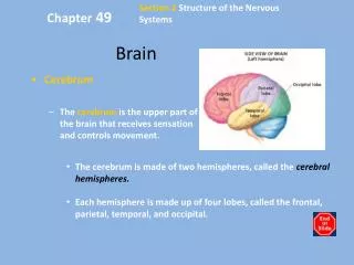
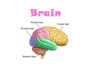
cerebrumcortex- gray/white matter • Rt/Lt hemispheres • Gyri- folds • Sulci- groove- central sulcus • frontal (motor) / parietal lobe (sensory) • Fissures- deeper grooves- • longitudnal- superior sagittal sinus/ flax (Rt/Lt) • lateral- Sylvian- frontal/ parietal from temporal lobe. • Corpus callosum- largest bundle of white matter w/ in cerebrum forms roof of the lateral vents • connects Rt/ Lt cerebral hemispheres • 4 parts= rostrum, genu, body, splenium
Corpus Callosum http://psycnet.apa.org/journals/neu/17/3/images/neu_17_3_496_fig1a.gif http://img.medscape.com/pi/emed/ckb/radiology/336139-407730-7910.jpg
Locate the corpus callosum… www.come-over.to/FAS/corpuscallosum3.jpg
What is wrong? http://neurosurgerydallas.com/images/2_1_3_5b.jpg
Label the arrows- 1,4, 6, 7… http://www.info-radiologie.ch/brain_mri_coronal_t2.php http://upload.wikimedia.org/wikipedia/commons/1/15/26638.medium-emphasizing-corpus-callosum.png
Cisterns • Subarachnoid space- areas at brain base that are widened where CSF pools. • Supracellar cistern- superior to sella- C of W • Quadrigeminal cistern- posterior to quadrigeminal plate of midbrain • Cisterna magna- lower posterior fossa (largest)
Cisterns http://pilgrimagetozion.files.wordpress.com/2009/07/cistern.jpg http://www.aans.org/bulletin/images/Vol17_2_08/Nonenhanced-axial-CT_large.jpg http://icanhascadherin.files.wordpress.com/2008/09/happy-face.jpg
What is wrong? http://www.scielo.br/img/revistas/anp/v66n3b/a32fig01.gif http://img.medscape.com/pi/emed/ckb/radiology/336139-336489-4514.jpg
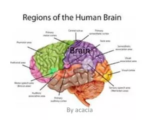
Cerebral Lobes • Cortex divided into 4 lobes: • Frontal- anterior- reason, judgment, vol muscle • Broca’s area for speech- LT frontal gyrus • Parietal- middle of each hemisphere- post to central sulcus- temperature, touch, pain, taste • Occipital- posterior- visual stimuli from thalamus • Temporal- anterior to occipital-auditory/olfactory • Primary auditory= Heschl’s- auditory info • Secondary= Wernicke’s – comprehension/ speech
Cerebral lobes http://space.newscientist.com/data/images/archive/2222/22224201.jpg http://www.ispub.com/ispub/ijra/volume_4_number_1_46/neurological_damage_in_heat_stroke_in_a_child_ct_mri_and_spect_appearances/heat-fig2b.jpg
temporal cerebellum http://www.imaios.com/var/ezwebin_site/storage/images/media/images/e-anatomy/brain-mri/en/brain-anatomy-axial-atlas/4651-1-eng-GB/brain-anatomy-axial-atlas_imagelarge.jpg http://www.nature.com/bmt/journal/v39/n4/images/1705571f1.jpg
Case study= Heat Stroke The described imaging findings in heat stroke include early cerebral edema , loss of gray-white matter differentiation, patchy high signal intensity of the white matter of cerebral hemispheres and vascular boundary zone infarcts and in later stages, diffuse cerebellar atrophy -severe brain ischemia was the underlying cause for the neurological involvement in heat stroke in our patient. http://images.google.com/imgres?imgurl=http://www.ispub.com/ispub/ijra/volume_4_number_1_46/neurological_damage_in_heat_stroke_in_a_child_ct_mri_and_spect_appearances/heat-fig2b.jpg&imgrefurl=http://www.ispub.com/journal/the_internet_journal_of_radiology/volume_4_number_1_46/article_printable/neurological_damage_in_heat_stroke_in_a_child_ct_mri_and_spect_appearances.html&usg=__MOWFvBWDRgjINv-DxcrF_mmMfwM=&h=575&w=477&sz=41&hl=en&start=3&itbs=1&tbnid=EXEYVfzs4fUVPM:&tbnh=134&tbnw=111&prev=/images%3Fq%3Dcerebral%2Bcortex%2Bon%2BMRI%2Bor%2BCT%26gbv%3D2%26hl%3Den%26sa%3DG
Basal Ganglia • Subcortical gray matter • Caudate nucleus- lat to each lat ventricle • Lentiform nucleus • Claustrum • Together they plan and program muscle action. http://www.brainexplorer.org/brain-images/brain_slice_small.jpg http://www.biomedcentral.com/content/figures/1471-2377-6-33-3-l.jpg
Diencephalon • Thalamus • Lg oval grey masses, walls of lat vents, connects in mid 3rd vent by massa intermedia • Hypothalamus • Inferior to thalamus and posterior to optic chiasm, making floor of lat vent- Pituitary Gland (hypophysis)- connected by infaundibulum. • Epithalamus • Pineal gland- melatonin= day/night cycles (can Ca++)
Limbic System • Interconnected fibers adjacent to temporal lobes. • Emotional aspects of behavior • Includes: hippocampus (short- long term memory converter), amygdala, olfactory tracts, fornix, cingulate gyrus.. • Brain injury to hippocampus can cause….. • Loss of memory…
Brainstem • Major segments are: • Midbrain • Above the pons, smallest portion of brainstem • Nerve bundles called cerebral peduncles and quadrigeminal plate. • Surrounds the cerebral aquaduct- which connects the 3rd and 4th vents and contains CSF • Pons • Oval expansion of brainstem- “Bridge”- signals to spinal cord and cerebral cortex • Medulla oblongata • Extends to the spinal cord which exits the foramen magnum- heart rate, respiratory rate and blood pressure.
Cerebellum • “Little brain” • Posterior to brainstem and occupies posterior fossa • Composed of two cerebellar hemispheres with a midline Vermis- on the inferior surface lie the cerebellar tonsils • Occassionally they may herniate down the foramen magnum…..called what????
Cerebellum Dandy Walker= refers to the enlargement of the posterior fossa secondary to cystic dilatation of the fourth ventricle as well as hypoplasia of the cerebellum and vermis.
What is wrong? Lt cerebellar abscess Congenital absence of one cerebellar hemisphere
Types of brain lesions… PNET-Primitive Neuroectodermal tumors PNET can occur anywhere in the brain of a child, although the most common place is in the back of the brain near the cerebellum. When they occur here, they are called medulloblastomas. The symptoms depend on their location in the brain, but typically the child experiences increased intracranial pressure. These tumors are fast growing and often malignant, with occasional spreading throughout the brain or spinal cord.
Cerebral Vasculature • Arteries in the brain are thin and weak. • high risk for aneurysms and strokes. • No valves in the veins-blood can flow in either direction= route for blood-bourne pathogens. • Dural sinus drains down to the IJ’s- through the superior and inferior sagittal sinus • Unique capillaries create the Blood Brain Barrier
Arterial- Blood to Brain • Paired internal carotid and vertebral arteries. • Internal carotid becomes- Carotid Siphon • Anterior cerebral-ACA • Middle cerebral-MCA • Circle of Willis= Located in Suprasellar cistern- cerebral arterial circle- anastomosis of 4 major arteries…. • What are they? • Two vertebral and Two carotids…. • becoming the anterior/posterior cerebral, anterior/posterior communicating and internal carotids
Venous Blood Parietal Veins Superior Sagital Sinus Confluence of Sinus Transverse Sinus Sigmoid Sinus Internal Jugular Veins
Which is it? MCA stroke- frontal/temporal lobe with Wedge shaped area of decreased density and Slight midline shift/ mass effect Aneurysm AVM
Cranial Nerves • 12 cranial • All but 1st and 2nd arise from the brainstem • Each nerve corresponds to a function in the body • Olfactory- smell- superior nasal septum • Optic-sight-posterior aspect of eye • Facial-lower pons- control facial muscles • Vagus-”wandering”- down to splenic flexure of abd and arise from medulla oblongata- enervates many abdominal organs