Trauma. Intra-Abdominal Injuries Crush Injuries Fracture . Dr. Huda B. Hassan
Trauma. Intra-Abdominal Injuries Crush Injuries Fracture . Dr. Huda B. Hassan. Emergency Approach to the Trauma Patient Injury is the fourth leading cause of death in the United States. Among patients aged 1-44 years, it is the leading cause of death.

Trauma. Intra-Abdominal Injuries Crush Injuries Fracture . Dr. Huda B. Hassan
E N D
Presentation Transcript
Trauma. Intra-Abdominal Injuries Crush Injuries Fracture. Dr. Huda B. Hassan
Emergency Approach to the Trauma Patient Injury is the fourth leading cause of death in the United States. Among patients aged 1-44 years, it is the leading cause of death. Trauma can be classified into blunt (e.g. vehicular, falls) or penetrating (e.g. gunshot wounds, stab wounds Death due to injury occurs in one of three time periods.
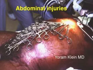
abdominal trauma A. Penetrating Abdominal Trauma Causes: -Gunshot wound -Stab wound -Embedded object from explosion Assessment: -Absence of bowel sound-Hypovolemic shock -Orthostatic hypotension-Pain and tenderness Management: 1. Maintain hemodynamic status –IVF & blood transfusion 2. Surgery-EXLAP 3. Peritoneal Lavage
Blunt Abdominal Trauma Assessment: -Left upper quadrant pain (Spleen) -Right upper quadrant pain (liver) -Signs of hypovolemic shock Management: 1. Maintain hemodynamic status 2. Monitor VS and oxygen supplements 3. Assess signs and symptoms of shock
Trimodal Death Distribution from Trauma: First Peak – occurs within seconds to minutes of injury Lacerations to the brain, brainstem, high spinal cord, heart, aorta Second Peak – occurs within minutes to hours of injury Subdural and epidural hematomas Hemopneumothorax Lacerated spleen or liver Pelvic fracture Other injuries associated with significant blood loss Third Peak – occurs several days to weeks after injury Sepsis Multi-system organ failure
it is essential to have a method of providing rapid patient assessment and interventions to decrease the number of deaths encountered in the second peak Triage Not all injured patients require transport to a Level I (highest level) trauma center. Criteria have been established to help pre-hospital personnel decide which patients to transport to such a center.
The composition of a trauma team is institution- Based Trauma resuscitations are “run” by a senior emergency medicine or senior surgical resident. Subspecialty consultants such as neurosurgeons and orthopedists are available on short notice. Referral to a trauma center is dependent on anatomic criteria and mechanism of injury criteria
Mechanism of Injury: • High-speed traffic crash (>40mph) • Vehicle space invaded > 20 inches • Pedestrian accident (age < 5 years or > 55 years) • Pedestrian accident (vehicle moving > 5mph) • Pedestrian thrown > 15 feet by car • Ejection from vehicle • Rollover • Death of other occupants of same vehicle • Prolonged extrication time (>20 minutes) • Falls greater than 20 feet
Anatomic factors: • Penetrating trauma to the head, neck, torso or groin to mid-thigh • Major burn (covering >20% partial-thickness or full-thickness skin area) • Major amputation above ankle or wrist • Limb paralysis • Two or more proximal long bone fractures • Flail chest, • Hemo- or pneumothoraces • Abnormality in vital signs • Abnormal or change of mental status in route • Significant patient comorbidities
Initial ED Resuscitation Assessments based on the primary and secondary surveys. Three important underlying concepts taught by ATLS are: • Treat the greatest threat to life first. Injury kills in reproducible time frames. The mnemonic ضع في ذاكرتك ABCDE was developed to order our evaluations and treatment such that most lethal problem is treated first. • The lack of a definitive diagnosis should not impede treatment. • A detailed history is not essential to begin the evaluation of the acutely injured patient.
Primary Survey • A – airway and C-spine stabilization • B – breathing • C – circulation • D – disability • E – exposure and environmental control
Airway is assessed first for patency and measures to establish a patent airway are instituted. • Methods for managing the airway include simple maneuvers such as chin lift, placement of a nasal airway (should not be used if there are facial injuries) or oropharyngealفموي بلعومي airway. • If these measures fail, bag-valve-mask ventilation should be attempted.
Breathing is assessed by visualizing movement of the chest and auscultating for breath sounds which should be equal and audible bilaterally. If there are signs of a hemo- or pneumothorax, a tension pneumothorax, open chest wounds or flail chest, then attention to these matters should be paid at this time. Tube thoracostomy should be done for hemothorax or pneumothorax
Circulation is assessed next, by evaluating for the presence of a pulse, heart tones, capillary refill, and warmth of the extremities. Although the blood pressure is not part of the ATLS algorithm, it is an important indicator of a patient’s hemodynamic status Initial fluid resuscitation is with 20ml/kg, of Lactated Ringer’s or normal saline. If vital signs are not improved after 30ml/kg (2 liters) of crystalloids, blood should be transfused. If cross matched blood is not available when the decision to transfuse is made, then type O blood should be given to the unstable patient.
Assessment of the circulatory status of the trauma patient includes evaluation for evidence of external hemorrhage There are five potential areas of blood loss that can lead to hypotension: • chest • abdomen/pelvis • Retro-peritoneum • thigh • on the street (external hemorrhage)
Disability, referring to the neurologic assessment, is evaluated next. A rapid means of evaluating neurologic status is the use of the AVPU mnemonic. The Glasgow Coma Scale is a more detailed method of evaluation and can be done as part of the secondary survey. AVPU A – alert V – responds to verbal stimuli, P – responds only to painful stimuli, U – unresponsive to all stimuli
Glasgow Coma Scale Score = (E+V+M) Best possible score = 15 Worst possible score = 3 Coma is defined as GCS less than or equal to 8: Severe head injury GCS 8 or below Moderate head injury GCS 8-13 Minor head injury GCS 14-15
Exposure of the trauma patient is essential for a thorough examination In order to obtain such exposure without moving the patient’s c-spine and/or injured limbs, it is often necessary to cut off the patient’s clothing. Once assessment is completed, the patient should be covered with warm blankets in order to prevent hypothermia
Other interventions are undertaken during the primary survey. These are called adjuncts اضافي to the primary survey and include: ECG monitoring, pulse oximetry, monitoring of vital signs, placement of a gastric tube to decompress the stomach in order to help decrease the risk of aspiration, and the placement of a Foley catheter for monitoring of urinary output.
. A nasogastric catheter is contraindicated in a patient with facial fractures, bleeding from the nostrils and CSF rhinorrhea, as the tube may penetrate the brain in patients with suspected cribriform plate injuries A Foley catheter should not be placed if there is suspicion of a urethral injury heralded by: 1) blood at the penile meatus, 2) perineal ecchymoses, 3) scrotal hematoma, 4) high-riding prostate or nonpalpable prostate, or 5) a pelvic fracture.
Remember, placement of gastric and urinary catheters is not a life-preserving intervention X-rays may be obtained. Examine the chest X-ray for injury to the bony structures such as the humerus, clavicles, ribs or scapula; correct placement of all lines and tubes; air in the soft tissues; free air under the diaphragm as an indicator of abdominal viscus perforation; pneumo- or hemothoraces; pulmonary contusions; and signs of aortic injury
Secondary Survey The secondary survey comprises a head-to-toe evaluation of the trauma patient and reevaluation of the vital signs. A focused history is obtained at this point using the AMPLE mnemonic. AMPLE A – allergies M – medications P – past illnesses/pregnancy L – last meal consumed E – events leading up to the injury
CT scan evaluation of the abdomen in the trauma patient has several benefits
Orthopaedic Injuries • Goals of ED Treatment • identify injuries accurately and address potentially life/limb threatening problems appropriately • reduce immobilize fractures (cast/splint) as appropriate • provide adequate pain relief • arrange proper follow-up if necessary
Physical Examination Rotation • Look (inspection): "SEADS" Swelling, Erythema, Atrophy, Deformity, Skin changes Translation (e.g. bruises) Alignment/angulation • Feel (palpation): all joints/bones -local tenderness, swelling, warmth, crepitus, joint Type (i.e. Salter-Harris, etc.) effusions, subtle deformity • Move: joints affected plus above and below injury -active ROM preferred to passive • Neurovascular status: distal to injury (BEFORE and AFTER reduction)
life and limb threatening injuries • threat to life is usually due to blood loss (e.g. up to 3 L in pelvic fractures, 1.5 L per long bone fracture) • threat to limb is usually due to interruption of blood supply to distal part of limb or to susceptible part of bone
•Open Fractures • communication between fracture site and external surface of skin -risk of osteomyelitis • remove gross debris, irrigate, cover with sterile dressing -formal irrigation and debridement often done in the OR • control bleeding with pressure (no clamping) • splint • antibiotics (15' generation cephalosporin and amino-glycoside) and tetanus prophylaxis • must secure definitive surgical care within 6-8 hours
Soft Tissue Injuries/ Emergency Wound Management • Goals of ED Treatment • identify injuries and stop any active bleeding -direct pressure • manage pain • wound examination and exploration (history and physical) • cleansing, ± antibiotic and tetanus prophylaxis • repair and dressing
Bruises • tender swelling (hematoma) following blunt trauma • is patient on anticoagulants? do they have a coagulopathy (e.g. liver disease)? • Abrasions • partial to full thickness break in skin • • management • dean thoroughly, ±. local anesthetic, with brush to prevent foreign body impregnation (tattooing) • antiseptic ointment (Polysporin™ or Vaseline™) for 7 days for facial and complex abrasions • tetanus prophylaxis -see Table 9 above
Lacerations • consider every structure deep to a laceration injured until proven otherwise • in hand injury patient, include following in history: handedness, occupation, mechanism of injury, previous history of injury • physical exam • think about underlying anatomy • examine tendon function actively against resistance and neurovascular status distally • clean and explore under local anesthetic; look for partial tendon injuries • x-ray wounds if a foreign body is suspected (e.g. shattered glass) and not found when exploring wound (remember: not all foreign bodies are radiopaque), or if suspect intra-articular involvement
• management • disinfect skin/use sterile techniques • irrigate copiously with normal saline • analgesia ± anesthesia • • maximum dose of lidocaine: • 7 mglkg with epinephrine • 5 mglkg without epinephrine
in children, topical anesthetics such as LET (lidocaine, epinephrine and tetracaine) and in selected cases a short-acting benzodiazepine (midazolam or other agents) for sedation and amnesia are useful • secure hemostasis • evacuate hematomas, debride non-viable tissue, remove hair and remove foreign bodies • • ± prophylactic antibiotics • suture unless delayed presentation, a puncture wound, or mammalian bite • take into account patient and wound factors when considering suturing • advise patient when to have sutures removed
Thank you for Listening Dr.Huda