An Introduction to Functional MRI
990 likes | 2.15k Vues
An Introduction to Functional MRI. Brain Imaging and Analysis Center FMRI Graduate Course. Summary of the Course. Combines lectures and laboratory sessions Laboratories will illustrate concepts from lectures Grading basis Participation in course sessions (attendance, discussion)

An Introduction to Functional MRI
E N D
Presentation Transcript
An Introduction to Functional MRI Brain Imaging and Analysis Center FMRI Graduate Course
Summary of the Course • Combines lectures and laboratory sessions • Laboratories will illustrate concepts from lectures • Grading basis • Participation in course sessions (attendance, discussion) • Completion of laboratory exercises • One take-home test (mid-term) • Practicum research project at end of semester • Course web page (www.biac.duke.edu/education) • BIAC Logins • Readings • Buxton, Introduction to fMRI • Original papers (generally posted on the web page) • Full reading list to be posted over upcoming weeks
Outline for Today • Why use fMRI to image brain function? • Key concepts of fMRI • History of fMRI • Parts of a MR scanner • MR safety • Laboratory: Scanner Visit (Dr. Jim Voyvodic) • Scanner hardware • Stimulus presentation and recording hardware • Demonstration of real-time fMRI
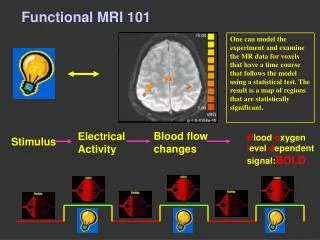
What is fMRI? • A technique for measuring changes in brain activity over time using principles of magnetic resonance. • Scanning procedures and restrictions are generally similar to clinical MR. • Most fMRI studies use changes in BOLD contrast, although other measures exist.
Why the Growth of fMRI? • Powerful • Improved ability to understand cognition • Better spatial resolution than PET • Allows new forms of analysis • High benefit/risk ratio • Non-invasive (no contrast agents) • Repeated studies (multisession, longitudinal) • Accessible • Uses clinically prevalent equipment • No isotopes required • Little special training for personnel
New Cognitive Analyses • Sampling rate affects experimental design • PET: >30s/data point ; fMRI: 1s/data point • Cognitive processes being measured must change more slowly than sampling rate • New forms of analyses • Event-related: sorting trials by accuracy, response time, type of condition • Rapid stimulus presentation • Allows creation of process models of activity • Difference in activation timing between regions is often on order of 100-1000ms
Cheng, Waggoner, & Tanaka (2001) Neuron Sakai, Rowe, & Passingham (2002) Nature Neuroscience
Key Concepts • Contrast • Spatial Resolution • Temporal Resolution • Functional Resolution
Anatomical Contrast Definition: The ability to distinguish between two (or more) different properties of tissue.
Blood Oxygenation Level Dependent (BOLD) Contrast From Mosley & Glover (1995)
Design Effects on Functional Contrast Contrast should really be considered as “contrast to noise”: how effectively can we decide whether a given brain region has property X or property Y?
Spatial Resolution: Voxels Voxel: A small rectangular prism that is the basic sampling unit of fMRI. Typical functional voxel: (4mm)3. Typical anatomical voxel: (1.5mm)3.
~8mm2 ~4mm2 ~2mm2 ~1.5mm2 ~1mm2 Spatial Resolution: Examples
Temporal Resolution • Importance depends upon research question • Type I: Detection • Temporal resolution is only indirectly important if your study investigates whether or not a given brain region is active. • Type II: Estimation • Temporal resolution is extremely important when attempting to understand the properties of an active region. • Determining factors • Sampling rate, usually repetition time (TR) • Dependent variable, usually BOLD response • BOLD response is sluggish, taking 2-3 seconds to rise above baseline and 4-6 seconds to peak • Experimental design
Functional Resolution The ability of a measurement technique to identify the relation between underlying neuronal activity and a cognitive or behavioral phenomenon. Functional resolution is limited both by the intrinsic properties of our brain measure and by our ability to manipulate the experimental design to allow variation in the phenomenon of interest.
1920 1930 1940 1950 1960 1970 1980 1990 2000 Timeline of MR Imaging 1972 – Damadian patents idea for large NMR scanner to detect malignant tissue. 1924 - Pauli suggests that nuclear particles may have angular momentum (spin). 1937 – Rabi measures magnetic moment of nucleus. Coins “magnetic resonance”. 1985 – Insurance reimbursements for MRI exams begin. 1973 – Lauterbur publishesmethod for generating images using NMR gradients. MRI scanners become clinically prevalent. 1944 – Rabi wins Nobel prize in Physics. 1952 – Purcell and Bloch share Nobel prize in Physics. NMR becomes MRI 1973 – Mansfield independently publishes gradient approach to MR. 1990 – Ogawa and colleagues create functional images using endogenous, blood-oxygenation contrast. 1946 – Purcell shows that matter absorbs energy at a resonant frequency. 1959 – Singer measures blood flow using NMR (in mice). 1975 – Ernst develops 2D-Fourier transform for MR. 1946 – Bloch demonstrates that nuclear precession can be measured in detector coils.
Discovery of Nuclear Magnetic Resonance Absorption (1946) • Bloch and Purcell independently discovered how to measure nuclear moment in bulk matter (1946) • Determined relaxation times. • They showed that energy applied at a resonant frequency was absorbed by matter, and the re-emission could be measured in detector coils • They shared the 1952 Nobel Prize in Physics Felix Bloch Edward Purcell
1920 1930 1940 1950 1960 1970 1980 1990 2000 Timeline of MR Imaging 1972 – Damadian patents idea for large NMR scanner to detect malignant tissue. 1924 - Pauli suggests that nuclear particles may have angular momentum (spin). 1937 – Rabi measures magnetic moment of nucleus. Coins “magnetic resonance”. 1985 – Insurance reimbursements for MRI exams begin. 1973 – Lauterbur publishesmethod for generating images using NMR gradients. MRI scanners become clinically prevalent. 1944 – Rabi wins Nobel prize in Physics. 1952 – Purcell and Bloch share Nobel prize in Physics. NMR becomes MRI 1973 – Mansfield independently publishes gradient approach to MR. 1990 – Ogawa and colleagues create functional images using endogenous, blood-oxygenation contrast. 1946 – Purcell shows that matter absorbs energy at a resonant frequency. 1959 – Singer measures blood flow using NMR (in mice). 1975 – Ernst develops 2D-Fourier transform for MR. 1946 – Bloch demonstrates that nuclear precession can be measured in detector coils.
Early Uses of NMR • Most early NMR was used for chemical analysis • No medical applications • 1971 – Damadian publishes and patents idea for using NMR to distinguish healthy and malignant tissues • “Tumor detection by nuclear magnetic resonance”, Science • Proposes using differences in relaxation times • No image formation method proposed • 1973 – Lauterbur describes projection method for creating NMR images • Mansfield (1973) independently describes similar approach
The First ZMR NMR Image Lauterbur, P.C. (1973). Image formation by induced local interaction: Examples employing nuclear magnetic resonance. Nature, 242, 190-191.
1920 1930 1940 1950 1960 1970 1980 1990 2000 Timeline of MR Imaging 1972 – Damadian patents idea for large NMR scanner to detect malignant tissue. 1924 - Pauli suggests that nuclear particles may have angular momentum (spin). 1937 – Rabi measures magnetic moment of nucleus. Coins “magnetic resonance”. 1985 – Insurance reimbursements for MRI exams begin. 1973 – Lauterbur publishesmethod for generating images using NMR gradients. MRI scanners become clinically prevalent. 1944 – Rabi wins Nobel prize in Physics. 1952 – Purcell and Bloch share Nobel prize in Physics. NMR becomes MRI 1973 – Mansfield independently publishes gradient approach to MR. 1990 – Ogawa and colleagues create functional images using endogenous, blood-oxygenation contrast. 1946 – Purcell shows that matter absorbs energy at a resonant frequency. 1959 – Singer measures blood flow using NMR (in mice). 1975 – Ernst develops 2D-Fourier transform for MR. 1946 – Bloch demonstrates that nuclear precession can be measured in detector coils.
Using MRI to Study Brain Function Somatosensory Cortex: Hammeke, et al., 1994 Visual Cortex: Kwong, et al., 1994
Parts of a MR Scanner BIAC 1.5T Scanner
MRI Safety Issue: The appropriate risk level for a research participant is much lower than for a clinical patient.
Hospital Nightmare Boy, 6, Killed in Freak MRI AccidentJuly 31, 2001 — A 6-year-old boy died after undergoing an MRI exam at a New York-area hospital when the machine's powerful magnetic field jerked a metal oxygen tank across the room, crushing the child's head. … ABCNews.com
MR Incidents • Pacemaker malfunctions leading to death • At least 5 as of 1998 (Schenck, JMRI, 2001) • E.g., in 2001 an elderly man died in Australia after being twice asked if he had a pacemaker • Blinding due to movements of metal in the eye • At least two incidents (1985, 1990) • Dislodgment of aneurysm clip (1992) • Projectile injuries (most common incident type) • Injuries (e.g., cranial fractures) from oxygen canister (1991, 2001) • Scissors hit patient in head, causing wounds (1993) • Gun pulled out of policeman’s hand, hitting wall and firing • Rochester, NY (2000)
Issues in MR Safety • Magnetic Field Effects • Known acute risks • Projectiles, rapid field changes, RF heating, claustrophobia, acoustic noise, etc. • Potential risks • Current induction in tissue at high fields • Changes in the developing brain • Epidemiological studies of chronic risks • Extended exposure to magnetic fields • Difficulty in assessing subjective experience • In one study, 45% of subjects exposed to a 4T scanner reported unusual sensations (Erhard et al., 1995)
Possible Effects of Magnetic Fields • Physiological • Red blood cells (especially sickled) may alter shape in a magnetic field • Some photoreceptors may align with the field. • Sensory (generally reported in high-field) • Nausea • Vertigo • Metallic taste • Magnetophosphenes
Risks of MRI • Projectile Effects: External • Projectile Effects: Internal • Radiofrequency Energy • Gradient field changes • Claustrophobia • Acoustic Noise • Quenching
Projectile Effects: External Chaljub (2001) Schenck (1996) “Large ferromagnetic objects that were reported as having been drawn into the MR equipment include a defibrillator, a wheelchair, a respirator, ankle weights, an IV pole, a tool box, sand bags containing metal filings, a vacuum cleaner, and mop buckets.” -Chaljub et al., (2001) AJR Chaljub (2001)
Radiofrequency Energy • Tissue Heating • Specific Absorption Rate (SAR; W/kg) • Pulse sequences are limited to cause less than a one-degree rise in core body temperature • Scanners can be operated at up to 4 W/kg (with large safety margin) for normal subjects, 1.5 W/kg for compromised patients (infants, fetuses, cardiac) • Weight of subject critical for SAR calculations • Burns • Looped wires can act as RF antennas and focus energy in a small area • Most common problem: ECG leads • Necklaces, earrings, piercings, pulse oximeters, any other cabling
Projectile/Torsion Effects: Internal • Motion of implanted medical devices • Clips, shunts, valves, etc. • Motion or rotation of debris, shrapnel, filings • Primary risk: Metal fragments in eyes • Swelling/irritation of skin due to motion of iron oxides in tattoo and makeup pigments
Acoustic Noise • Potential problem with all scans • Short-term and long-term effects • Sound level of BIAC scanners • 1.5T: 93-98 dB (EPI) • 4.0T: 94-98 dB (EPI) • OSHA maximum exposure guidelines • 2-4 hours per day at BIAC levels • Earplugs reduce these values by 14-29 dB, depending upon fit.
Gradient Field Changes • Peripheral nerve stimulation • May range from distracting to painful • Risk greatly increased by conductive loops • Arms clasped • Legs crossed • Theoretical risk of cardiac stimulation • No evidence for effects at gradient strengths used in MRI
Claustrophobia • Most common subject problem • About 10% of patients • About 1-2% of BIAC subjects • Ameliorated with comfort measures • Talking with subject • Air flow through scanner • Panic button • Slow entry into scanner
Quenching • Definition: Rapid decrease in magnetic field strength due to loss of superconductivity • Only initiated voluntarily due to danger to participant’s life or health • Effects • Magnets heat up with loss of current • Cryogenic fluids (Helium) boil off and fill the scanner room • Displaces breathable air from room • Cooling of room, condensation reduces visibility • Physical damage to the scanner may occur • Safety personnel must be cognizant of room conditions
Scanner Visit • Anyone with implanted metal should see me before going to the scanner • Pacemaker, cochlear implant, shunt, clip, etc. • Dental work and piercings are fine • Please do not bring backpacks, books, etc., • You can leave them here in the room, which we will lock • MR center is a working patient environment