Chapter 8: Functional MRI
Chapter 8: Functional MRI. CS689 Computational Medical Imaging analysis. Spring 2009 University of Kentucky. Outline. fMRI vs. MRI Procedures of fMRI Medical Significance of fMRI Methods of fMRI Types of fMRI. a Simple Definition:.

Chapter 8: Functional MRI
E N D
Presentation Transcript
Chapter 8: Functional MRI CS689 Computational Medical Imaging analysis Spring 2009 University of Kentucky
Outline • fMRI vs. MRI • Procedures of fMRI • Medical Significance of fMRI • Methods of fMRI • Types of fMRI 4/3/2007 University of Kentucky
a Simple Definition: If the MRI experiment is done while a mental task is given to a subject, a so-called functional magnetic resonance image (fMRI) is generated. fMRI is used to map different sensor, motor and cognitive functions to specific regions in the brain. 4/3/2007 University of Kentucky
fMRI vs. MRI MRI studies brain anatomy. Seeing brain structure 3Dbrain anatomy Functional MRI (fMRI) studies brain function. 4/3/2007 University of Kentucky
Procedures of fMRI • a series of baseline images are taken of the brain region of interest when the subject is at rest, as A • the subject performs a task • a second series of images is taken, as A’ • the first set of images is subtracted from the second, as B=A’-A • the areas that are most visible in the resulting image, B, are presumed to have been activated by the task. 4/3/2007 University of Kentucky
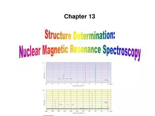
A healthy subject was asked to listen to sentences being spoken while watching a screen with a flashing checkerboard presented. The sentences started and stopped at slightly different times than the flashing picture was turned on and off. One example On the basis of the differences in timing activation in the brain, the areas responsible for hearing (in the middle of the brain in grey) and vision (in the back of the brain in white) could be localized by fMRI (Image courtesy of Dr. S. Smith from www.fmrib.ox.ac.uk). Visual cortex Auditory cortex 4/3/2007 University of Kentucky
a Experiment • Timing: • It consists of a set of trials, and the data is partitioned into trials. • For some of these intervals, the subject simply rested, or gazed at a fixation point on the screen. For other trials, the subject was shown a picture and a sentence, and instructed to press a button to indicate whether the sentence correctly described the picture. • For these trials, the sentence and picture were presented in sequence, with the picture presented first on half of the trials, and the sentence presented first on the other half of the trials. • Forty such trials are available for each subject. The timing within each such trial is as follows: 4/3/2007 University of Kentucky
The first stimulus (sentence or picture) was presented at the begining of the trail (image=1). • 4 seconds later (image=9) the stimulus was removed, replaced by a blank screen. • 4 seconds later (image=17) the second stimulus was presented. This remained on the screen for 4 seconds, or until the subject pressed the mouse button, whichever came first. • A rest period of 15 seconds (30 images) was added after the second stimulus was removed from the screen. Thus, each trial lasted a total of approximately 27 seconds (approximately 54 images). • Imaging parameters: Images were collected every 500msec. Only a fraction of the brain of each subject was imaged. The data is marked up with 25-30 anatomically defined regions (called "Regions of Interest", or ROIs). 4/3/2007 University of Kentucky
Medical Significance of fMRI • Different tasks activate different parts of the brain • When listening to music, a specialized area in the auditory cortex along the sides of the brain shows an increased signal • The locations vary for different cases and individuals. 4/3/2007 University of Kentucky
Medical Significance of fMRI • a diagnostic method • learning how a normal, diseased or injured brain is working • assessing the potential risks of surgery or other invasive treatment of the brain. • planning brain surgery -- monitor normal brain function as well as any disturbed brain function. • While research is still ongoing, it appears that fMRI can also help assess the effects of stroke, trauma or degenerative disease (such as Alzheimer's) on brain function. 4/3/2007 University of Kentucky
fMRI? • It aims to determine the neurobiological correlation of behavior by identifying the brain regions (or “functioning modules”) that become “active” during the performance of specific tasks in vivo. • It extends traditional anatomical imaging to functional imaging. • observe both the structures and which structures participate in specific functions • improve our understanding of a variety of brain pathologies. • such as the addictive behaviors of gambling or drug abuse, are without structural brain changes. 4/3/2007 University of Kentucky
the principle: • when a brain region is being used, arterial oxygenated blood will redistribute and increase to this area. 4/3/2007 University of Kentucky
Knowledge required • The science of applying fMRI is quite complicated and multi-disciplinary. It involves: • Physics: • Statistics: Because the signals are very subtle, correct application of statistics is essential in the statistical analysis of results to "tease out" observations and avoid false-positive results. • Psychology: When conducting fMRI on humans it is essential to employ carefully designed psychophysical experiments which allow the precise measurement of the neural effect under consideration. • Neuroscience: For a non-invasive scan, MRI has moderately good spatial resolution, but relatively poor temporal resolution. Increasingly, it is being combined with other data collection techniques such as electroencephalography (EEG) or magnetoencephalography (MEG), which have much higher recording frequencies. • Neuroanatomy: Anatomy is critical in understanding the location (and role) of the signals which fMRI is able to detect. 4/3/2007 University of Kentucky
3. Types of functional MRI: • BOLD fMRI • measures blood oxygenation, i.e, regional differences in oxygenated blood • perfusion fMRI • measures regional cerebral blood flow,i.e. the rate at which blood is delivered to tissue. • diffusion-weighted fMRI • measures random movement of water molecules in tissue. • It can detect acute brain infarction within 1 to 2 hours ***Infarct: 梗塞,如由于血栓或栓子的原因,局部血液供应不畅而引发局部组织坏死 • Magnetic Resonance Spectroscopic Imaging (MRSI) • measure certain cerebral metabolites noninvasively. • phase encoding is used to obtain spectra from multiple regions across a field of view. 4/3/2007 University of Kentucky
3.1 BOLD fMRI • Blood Oxygen Level Dependent Contrast • Based on: • (1) different magnetic properties of deoxy- and oxyhemoglobin (氧合血红蛋白) • (2) coupling of oxygenated blood flow and neuronal activity • High spatial and temporal resolution • 3-6 second delay in hemodynamic(血液动力学) response ---limits optimal temporal resolution. • Compares images taken during active and rest states within a single session 4/3/2007 University of Kentucky
MRI vs. fMRI MRI fMRI high resolution (1 mm) low resolution (~3 mm but can be better) one image • fMRI • Blood Oxygenation Level Dependent (BOLD) signal • indirect measure of neural activity … many images (e.g., every 2 sec for 5 mins) neural activity blood oxygen fMRI signal 4/3/2007 University of Kentucky
VOXEL (Volumetric Pixel) Slice Thickness e.g., 6 mm In-plane resolution e.g., 192 mm / 64 = 3 mm 3 mm 6 mm SAGITTAL SLICE IN-PLANE SLICE 3 mm Number of Slices e.g., 10 Matrix Size e.g., 64 x 64 Field of View (FOV) e.g., 19.2 cm Slice Terminology 4/3/2007 University of Kentucky
Hemoglobin Figure Source, Huettel, Song & McCarthy, 2004, Functional Magnetic Resonance Imaging 4/3/2007 University of Kentucky
Magnetic properties of deoxy- and oxyhaemoglobin • Deoxyhaemoglobin is paramagnetic • The presence of deoxyhaemoglobinin vessel causes a susceptibilitydifference between the vessel and its surrounding tissue. • Then causes dephasing of MR proton signal • Leading to a reduction in the value of T2*, which causes a darkening of the image • oxyhaemoglobin is diamagnetic 4/3/2007 University of Kentucky
Susceptibility artifacts • The magnetic susceptiblity of a material :::: a measure of how much magnetization is produced within it when it is placed in a magnetic field. • Susceptibility differences between tissues can lead to signal loss in MR scans, especially in EPI scans. • The susceptibility difference between deoxygenated and oxygenated blood is the basis of the BOLD effect used to detect fMRI signals. 4/3/2007 University of Kentucky
HDR (Hemodynamics Response) • the time courses of the changes in blood flow, blood volume and blood oxygenation that occur in the brain in response to brain activity. • In the brain, neuronal activity is thought to cause a local increase in blood flow (CBF), which leads to an increase in blood oxygenation and blood volume (CBV). • Upon activation, oxygen is extracted by the cells, increasing the level of deoxyhaemoglobin in the blood. And it is compensated for by an increase in blood flow in the vicinity of the active cells, leading to a net increase in oxyhaemoglobin. • Signal changes 4/3/2007 University of Kentucky
T2*-weighted imaging • T2*-weighted images are performed which take advantage of the different magnetic properties of deoxy- and oxyhaemoglobin. • Because of the magnetic properties of the deoxyhaemoglobin molecule which causes rapid dephasing, T2* signal is retained longer in a region when it has more oxygenated blood compared to when there is less oxygenated blood. • Thus, an area with more oxygenated blood will show up more intense on T2*-weighted images compared to when there is less oxygenated blood around. 4/3/2007 University of Kentucky
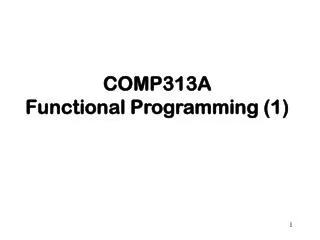
BOLD Time Course 4/3/2007 University of Kentucky
Evolution of BOLD Response Hu et al., 1997, MRM 4/3/2007 University of Kentucky
Initial Dip (Hypo-oxic Phase) • Transient increase in oxygen consumption, before change in blood flow • Menon et al., 1995; Hu, et al., 1997 • Smaller amplitude than main BOLD signal • 10% of peak amplitude (e.g., 0.1% signal change) • Potentially more spatially specific • Oxygen utilization may be more closely associated with neuronal activity than positive response Slide modified from Duke course 4/3/2007 University of Kentucky
Rise (Hyperoxic Phase) • Results from vasodilation of arterioles, resulting in a large increase in cerebral blood flow • Inflection point can be used to index onset of processing Slide modified from Duke course 4/3/2007 University of Kentucky
Peak – Overshoot • Over-compensatory response • More pronounced in BOLD signal measures than flow measures • Overshoot found in blocked designs with extended intervals • Signal saturates after ~10s of stimulation Slide modified from Duke course 4/3/2007 University of Kentucky
Sustained Response • Blocked design analyses rest upon presence of sustained response • Comparison of sustained activity vs. baseline • Statistically simple, powerful • Problems • Difficulty in identifying magnitude of activation • Little ability to describe form of hemodynamic response • May require detrending of raw time course Slide modified from Duke course 4/3/2007 University of Kentucky
Undershoot • Cerebral blood flow more locked to stimuli than cerebral blood volume • Increased blood volume with baseline flow leads to decrease in MR signal • More frequently observed for longer-duration stimuli (>10s) • Short duration stimuli may not evidence • May remain for 10s of seconds Slide modified from Duke course 4/3/2007 University of Kentucky
BOLD fMRI • BOLD = convolution of neuronal activity and haemodynamic transfer function (gamma) Neuronal Activity BOLD Signal Haemodynamic Function Time Time Slide from Matt Brown 4/3/2007 University of Kentucky
BOLD Summates Neuronal Activity BOLD Signal Slide from Matt Brown 4/3/2007 University of Kentucky
BOLD Overlap and Jittering • Closely-spaced haemodynamic impulses summate. • Constant ITI causes tetanus. Burock et al. 1998. 4/3/2007 University of Kentucky
Design Types = trial of one type (e.g., face image) = null trial (nothing happens) = trial of another type (e.g., place image) Block Design Slow ER Design Rapid Counterbalanced ER Design Rapid Jittered ER Design Mixed Design 4/3/2007 University of Kentucky
* Example • With prior knowledge of the activation timing, we can perform a statistical test on the data to determine which areas of the brain are active, then overlay this statistical map (shown in color) on a high resolution MR image so that one can visualize the functional information in relation to relevant anatomical landmarks. • There are a wide variety of different software packages that facilitate processing, analysis and display of fMRI data in addition to many different stimulus delivery packages: http://www.fmri-world.de/ The choice of each depends largely on the onsite resources and the specific application. A typical BOLD time course with 4 “active” states and 4 “resting” states. 4/3/2007 University of Kentucky
* Functional mapping : Statistical Parametric Mapping • Statistical parametric mapping entails the construction of spatially extended statistical processes to test hypotheses about regionally specific effects (Friston et al. 1991). • image processes with voxel values that are distributed according to a known probability density function, usually T or F distributions. 4/3/2007 University of Kentucky
General Linear Model (GLM) • To estimate some parameters that could explain the spatially continuous data in exactly the same way as in conventional analysis of discrete data. 4/3/2007 University of Kentucky
Gaussian random field • GRF theory is used to resolve the multiple-comparisons problem that ensues when making inferences over a volume of the brain. • GRF theory provides a method for adjusting p values for the search volume of an SPM to control false positive rates. 4/3/2007 University of Kentucky
Bayesian inferences • Bayesian inferences about spatially extended effects use posterior probability maps (PPMs). 4/3/2007 University of Kentucky
* Two issues on BOLD fMRI • No prior knowledge of activation timing • Temporal limits: HDR delay • Spatial limits of fMRI 4/3/2007 University of Kentucky
1. When the temporal response is unknown… • new fMRI analysis methods to detect activation without prior knowledge of activation timing. • One of these methods is called Independent Component Analysis (ICA) • It involves the development of novel ICA algorithms that are specific to fMRI data, the development of new stimulus designs that are appropriate for ICA and the use of ICA in patient populations to remove noise and motion artifacts. 4/3/2007 University of Kentucky
2. HDR delay & Limited temporal resolution • A time delay from the onset of the neural activity to the change of BOLD signal. • a time delay of 3-6 seconds between when a brain region is activated and blood flow increases to it. • During this time, the activated areas experience a relative decrease in oxygenated blood as oxygen is extracted by the active regional neurons. Afterward, the amount of blood flowing to the area far outweighs the amount of oxygen that is extracted so that oxygenated blood is now higher. 4/3/2007 University of Kentucky
Limited temporal resolution • Although images can be acquired every 100 msecs with EPI, this predictable but time-varied delayed onset of the BOLD response limits the immediate temporal resolution to several seconds instead of the 100 msec. • In the future, researchers may be able to improve the temporal resolution of fMRI by measuring the initial decrease in oxygenated blood with activation. 4/3/2007 University of Kentucky
3.What Limits Spatial Resolution • noise • smaller voxels have lower SNR • head motion • the smaller your voxels, the more contamination head motion induces • temporal resolution • the smaller your voxels, the longer it takes to acquire the same volume • 4 mm x 4 mm at 16 slices/secOR 1 mm x 1 mm at 1 slice/sec • vasculature • depends on pulse sequences • e.g., spin echo sequences reduce contributions from large vessels • some preprocessing techniques may reduce contribution of large vessels (Menon, 2002, MRM) 4/3/2007 University of Kentucky
fMRI for Dummies The Initial Dip • The initial dip seems to have better spatial specificity • However, it’s often called the “elusive initial dip” for a reason 4/3/2007 University of Kentucky
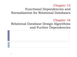
EXCEPT when the activated region does not fill the voxel (partial voluming) fMRI for Dummies Voxel Size non-isotropic non-isotropic isotropic 3 x 3 x 6 = 54 mm3 e.g., SNR = 100 3 x 3 x 3 = 27 mm3 e.g., SNR = 71 2.1 x 2.1 x 6 = 27 mm3 e.g., SNR = 71 In general, larger voxels buy you more SNR. 4/3/2007 University of Kentucky
fMRI for Dummies Partial Voluming • The fMRI signal occurs in gray matter (where the synapses and dendrites are) • If your voxel includes white matter (where the axons are), fluid, or space outside the brain, you effectively water down your signal 4/3/2007 University of Kentucky
fMRI for Dummies Partial Voluming Partial volume effects: The combination, within a single voxel, of signal contributions from two or more distinct tissue types or functional regions (Huettel, Song & McCarthy, 2004) This voxel contains mostly gray matter This voxel contains mostly white matter This voxel contains both gray and white matter. Even if neurons within the voxel are strongly activated, the signal may be washed out by the absence of activation in white matter. Partial voluming becomes more of a problem with larger voxel sizes Worst case scenario: A 22 cm x 22 cm x 22 cm voxel would contain the whole brain 4/3/2007 University of Kentucky
3.2. Diffusion and Perfusion MRI • Diffusion MRI measures the molecular mobility of water in tissue, while perfusion MRI measures the rate at which blood is delivered to tissue. • Therefore, both these techniques measure quantities which have direct physiological relevance. • Diffusion in biological systems is a complex phenomenon, influenced directly by tissue microstructure, and that its measurement can provide a large amount of information about the organization of this structure in normal and diseased tissue. • Perfusion reflects the delivery of essential nutrients to tissue, and so is directly related to its status. 4/3/2007 University of Kentucky
3.3. MRS • Magnetic Resonance Spectroscopy. The data gathered in MRS is presented as a spectrum. (i.e., the strength of the magnetic resonance signal is plotted as a function of resonant frequency). • Because of the way magnetic resonance works, the chemical environment of the nucleus being scanned will vary its resonant frequency. Hence, by observing the position of peaks in MR spectroscopic data it is possible to determine some of the molecules present in the sample. 4/3/2007 University of Kentucky