Exploring Cell Structures: Microscopes & Characteristics
Delve into the world of cell structure, from the 1600s discoveries to modern microscopy techniques. Learn about cell sizes, common features, and differences between prokaryotic and eukaryotic cells. Uncover the role of the cytoskeleton and the cell membrane in cellular function.

Exploring Cell Structures: Microscopes & Characteristics
E N D
Presentation Transcript
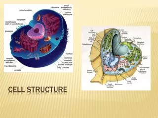
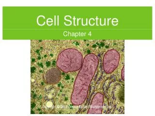
Cell Structure Chapter 3
3.1 Looking at Cells • 1600’s the microscope was invented • Robert Hooke 1665 looked at cork and saw little boxes he called “cells”
Anton Van Leeuwenhoek later looked at pond water and saw tiny animals he called “animalcules”
Measuring Cell Structures • Based on metric system giga- mega- kilo- hecto- deka- BASE deci- centi- milli- micro- nano- Giga = G Mega = M Micro = µ Nano = n
Characteristics of Microscopes • Light Microscope – light passes through 1 or more lenses • Electron Microscope – image made by beams of electrons • Micrograph – image from a microscope • Labeled with what kind of microscope and magnification value • Magnification – how many times larger it appears • Resolution – measure of clarity Electron Micrograph Ebola Virus 160,000 x magnification
Types of Microscopes 40 x • Compound Light Microscope • 2 lenses with light bulb shining through slide • Objective lens in close to slide • Ocular lens is near eye • 40x x 10x = 400x magnification • Third lens hurts resolution • Most powerful = 2,000x • See something 0.5 µm in diameter 400 x
Electron Microscope • Up to 200,000x • Beam and specimen must be in vacuum so e- don’t bounce off gas (no living things) • Transmission Electron Microscope • Stained with metal ions • Very thin slices of specimen • Internal structures • Black and white but computers add color • Scanning Electron Microscope • Specimen coated with thin layer of metal • 3-D cell surface • Artificial color
Scanning Tunneling Microscope • Needlelike probe measures differences in voltage caused by e- that tunnel from surface of object • 3-D image • Used on living things
3.2 Cell Features • Cell Theory • All living things are made of cells • Cells are the basic units of structure and function in organisms • All cells arise from existing cells
Cell Size • Smaller is more efficient • Everything must cross cells surface • Surface area to volume ratio • Too low - substances cannot enter and leave in large enough numbers • Small cells have a high ratio
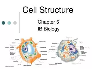
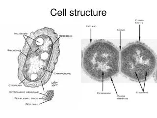
Common Features of Cells • Cell Membrane • Outer boundary enclosing cell that separates interior • Controls flow in and out • Cytoplasm • Cell interior
Cytoskeleton • System of microscopic fibers • Act as support structures
Ribosomes • Where proteins are made • DNA • All cells have it • Protein instructions • Regulate cellular activities • Allows for reproduction
Prokaryotes • Smallest, simplest, single-celled organism • No nucleus (and other parts missing) • Cannot carry out many special functions due to missing parts • Around at least 3.5 billion years ago (bya) • Nearly 2 bya were only organisms on earth • Very small from 1 – 15 µm • Bacteria • Subset that causes infection and food spoiling
Characteristics • Large range of environments • Many grow and divide rapidly • Some don’t need oxygen, others can’t have it • Some make own food
Everything inside cell membrane is in cytoplasm • Enzymes and ribosomes free to move around • Singular, circular DNA located in center • Cell Wall – surround membrane • Provides structure and support • No cytoskeleton so cell wall gives shape • Cell wall made of polysaccharides connected by short amino acid chains • Some cell walls surrounded by capsule = polysaccharides (allows them to cling to many things)
Flagella – long, threadlike structures that provide movement
Eukaryotic Cells • Internal compartments • Evolved about 2.5 bya • Have an internal compartment for DNA = nucleus • Have structures that carry out specific activities = organelles • Cytoplasm = everything inside membrane but outside nucleus • Membranes connect organelles and provide channels • Form envelopes called vesicles that move proteins between organelles
Flagella • Cilia – short, hair-like structures • Used for cell movement or movement of materials over cell • Cytoskeleton • Protein fibers • Holds cell together, keeps it from collapsing • Cytosol – fluid surrounding organelles, internal membranes and cytoskeleton
The Cytoskeleton • Interior framework of animal cell • Protein fibers anchored to inside of plasma membrane • 3 kinds of fibers • Actin Fibers • Long, slender microfilaments • Made of the protein actin • Determine shape of animal cells • Microtubules • Hollow tubes • Made of protein tubulin • Highway for transport from nucleus to parts of cell • Intermediate fibers • Thick ropes of protein • Frame for ribosomes and enzymes keeping them in certain locations
The Cell Membrane • Fluid, like a soap bubble • Lipids form a barrier allowing only certain things through - selective permeability • Phospholipid = 2 fatty acids and a phosphate group • Polar head – phosphate group; attracted to H20 • 2 nonpolar fatty acid tails; repelled by H20 • Phospholipids are in a double layer so called the lipid bilayer • Allows lipids and nonpolar substances to pass through
Membrane Proteins • Proteins are made of amino acids • Some are polar and some are nonpolar • Some move around • Marker proteins attached to carbohydrate tell what kind of cell it is (liver, heart) • Receptor proteins bind specific substances (signal molecules) • Enzymes in cell membrane important in biochemical reactions • Transport Proteins help move things in and out
3.3 Cell Organelles • Nucleus • Controls most cell functions • Surrounded by nuclear envelope • Double membrane (two lipid bilayers) • Nuclear pores = small channels allow passage through • Nucleolus – ribosomes partially assembled here • Most DNA stored here • Wound around proteins but in elongated thin strands • When about to divide they become more compact into chromosomes and form dense rod shaped structures • Number of chromosomes depends on species • Humans = 46 • Peas = 14
Ribosomes and Endoplasmic Reticulum • Ribososmes • Where proteins are made • Made of dozens of different proteins and RNA • May be free in cytosol but proteins made there stay in cell
Production of Proteins • Endoplasmic Reticulum is a system of internal membranes that move proteins and other structures through the cell • It is a lipid bilayer with embedded proteins • Rough ER • Attached ribosomes • Proteins made enter ER • Pinched off and form vesicles • Keep separate • Smooth ER • No ribosomes • Make lipids • Break down toxic substances
Packaging and Distribution of Proteins • Vesicles go from ER to Golgi Apparatus • Golgi Apparatus • Flattened membrane-bound sacs • Enzymes inside modify proteins • Modified proteins repackaged by GA and bud off • Lysosomes – vesicle that contains digestive enzymes
Steps of Protein packaging and distribution • Ribosomes on Rough ER make proteins and they are packaged • Go from ER to GA • In the GA proteins are modified and repackaged • Many vesicles move to cell membrane and release cargo • Other vesicles remain in cell and go a job
Mitochondria • Harvests energy from organic compounds to make ATP which is used as energy by cell • Most ATP is made here • Cells that use ATP have lots of mitochondria • 2 membranes • Outer = smooth • Inner = folded; large surface area • Forms 2 compartments
Mitochondrial DNA • DNA and ribosomes make their own proteins though most come from cytosol • Mitochondrial DNA (mDNA) is independent from nuclear DNA, similar to circular DNA of prokaryotic cell • Believe prokaryotes were ancestors to mitochondria
Structure of Plant Cells • Cell Wall • Surrounds cell membrane • Made of proteins and carbohydrates (cellulose) • Helps support and maintain shape • Protection • Connects to other cells
Chloroplasts • Use light to make carbohydrates from CO2 and H20 • Along with mitochondria supplies much of energy needed to power cell • 2 membranes • Contain their own DNA (ancient prokaryotes)
Central Vacuole • Stores water • May contain ions, nutrients, or wastes • When full, cell is rigid