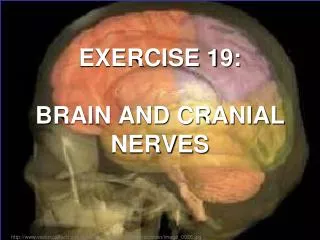
Exercise 19
Exercise 19. Spinal Cord & Spinal Nerves. http://www.georgiapainphysicians.com/downloads/m1_slides/4.%20Spinal%20cord%20junctions.jpg. Figure 19.1a Gross structure of the spinal cord, dorsal view. Spinal Cord. Extends from Foramen magnum L1-L2 @ conus medullaris. Cervical

Exercise 19
E N D
Presentation Transcript
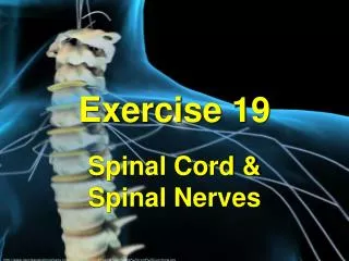
Exercise 19 Spinal Cord & Spinal Nerves http://www.georgiapainphysicians.com/downloads/m1_slides/4.%20Spinal%20cord%20junctions.jpg
Figure 19.1a Gross structure of the spinal cord, dorsal view. Spinal Cord Extends from Foramen magnum L1-L2 @ conus medullaris Cervical spinal nerves C1-C8 Thoracic spinal nerves T1- T12 Cauda equina “horse’s tail” lumbar & sacral nerves’ long ventral & dorsal roots… Filumterminale Fibrous extension of meninges, blends into coccygeal ligament Attaches to coccyx Conus medullaris Cauda equina Lumbar spinal nerves L1- L5 Filum terminale Sacral spinal nerves S1- S5 Coccygeal spinal nerve Co1 The spinal cord and its nerve roots, with the bony vertebral arches removed. The dura mater and arachnoid mater are cut open and reflected laterally.
Figure 19.1a Gross structure of the spinal cord, dorsal view. Medulla oblongata (brainstem) Conusmedullaris tapered inferior end Cervical spinal nerves C1-C8 Thoracic spinal nerves T1- T12 Conus medullaris Cauda equina Lumbar spinal nerves L1- L5 Filum terminale Sacral spinal nerves S1- S5 Coccygeal spinal nerve Co1 The spinal cord and its nerve roots, with the bony vertebral arches removed. The dura mater and arachnoid mater are cut open and reflected laterally.
Spinal Cord—Similar to Brain Gray matter Central in spinal cord White matter External in spinal cord Cerebrospinal Fluid (CSF) Fills meninges Proteins, nutrients—continuous circulation Pia mater Arachnoid mater Spinal dura mater • Meninges: surrounding membranes • Pia mater: innermost • Arachnoid: middle • Dura mater: outermost • Extend beyond the spinal cord • Continuous with meninges of brain • Physical stability, shock absorption
Figure 19.2a Anatomy of the human spinal cord. Spinal Cord—Similar to Brain Epidural space (contains fat) Pia mater Arachnoid mater Spinal meninges Dura mater (contains CSF) Bone of vertebra Dorsal root ganglion Spinal nerve Body of vertebra • Epidural space • between dura mater & walls of vertebrae • areolar tissue, blood vessels, adipose tissue (protection)
Spinal Nerves Figure 19.5 Human spinal nerves. Cervical plexus C1 – C5 Cervical spinal nerves Thoracic spinal nerves Lumbar spinal nerves Sacral spinal nerves Cervical nerves C1 –C8 Brachial plexus C5 – T1 Cervical enlargement Intercostal nerves Thoracic nerves T1 –T12 Lumbar enlargement Lumbar nerves L1 –L5 Lumbar plexus L1 – L4 Sacral plexus L4 – S4 Sacral nerves S1 –S5 Cauda equina Coccygeal nerve Co1
Nerves for Divisions • Sympathetic division – • thoracic & lumbar spinal nerves (thoracolumbar) • Parasympathetic division – • cranial nerves & sacral spinal nerves (craniosacral);
Spinal Nerve Plexuses Cervical nerves Thoracic nerves Lumbar nerves Sacral nerves Cervical plexus Brachial plexus Intercostal nerves (no plexus) Lumbar plexus Sacral plexus
Figure 19.6 The cervical plexus. Ventral rami Segmental branches Hypoglossal nerve (XII) Ventral rami: Lesser occipital nerve C1 Greater auricular nerve C2 Transverse cervical nerve C3 C4 Ansa cervicalis Accessory nerve (XI) C5 Phrenic nerve Supraclavicular nerves
Figure 19.7a The brachial plexus. Roots (ventral rami): Dorsal scapular Anterior divisions C4 Nerve to subclavius C5 Posterior divisions Suprascapular C6 Trunks Upper Posterior divisions C7 Middle Trunks Roots Lateral C8 Lower Cords T1 Posterior Long thoracic Medial Medial pectoral Lateral pectoral Axillary Upper subscapular Musculo- cutaneous Lower subscapular Radial Thoracodorsal Median Medial cutaneous nerves of the arm and forearm Ulnar Roots (rami C5 T1), trunks, divisions, and cords
Figure 19.7c The brachial plexus. Anterior divisions Axillary nerve Posterior divisions Trunks Roots Lateral cord Musculocutaneous nerve Posterior cord Medial cord Axillary nerve Humerus Radial nerve Radial nerve Median nerve Biceps brachii Musculo- cutaneous nerve Ulnar nerve Ulna Radius Cadaver photo Ulnar nerve Median nerve Radial nerve (superficial branch) Dorsal branch of ulnar nerve Superficial branch of ulnar nerve Digital branch of ulnar nerve Muscular branch Median nerve Digital branch The major nerves of the upper limb
Figure 19.8 The lumbar plexus (anterior view.) Ventral rami Ventral rami: L1 Iliohypogastric Ilioinguinal L2 Femoral Iliohypogastric Lateral femoral cutaneous Ilioinguinal L3 Genitofemoral Obturator Lateral femoral cutaneous Anterior femoral cutaneous L4 Obturator Saphenous L5 Femoral Lumbosacral trunk
Figure 19.9b The sacral plexus (posterior view). Ventral rami Superior gluteal Inferior gluteal Pudendal Sciatic Posterior femoral cutaneous Common fibular Tibial Sural (cut) Deep fibular Superficial fibular Plantar branches
Sympathetic Chain Ganglia • http://www.youtube.com/watch?v=fANkXK43xqk
Ganglia for Divisions • sympathetic ganglia – • along spinal column (proximal to CNS) • parasympathetic ganglia – • in or near the organs they control (distal to CNS)
Figure 19.2b Anatomy of the human spinal cord. Gray commissure Dorsal funiculus Dorsal horn White columns Ventral funiculus Gray matter Ventral horn Lateral funiculus Lateral horn Dorsal root ganglion Central canal Spinal nerve Dorsal root Pia mater Arachnoid mater Ventral root Spinal dura mater
Spinal Cord Anatomy Each segment has: Dorsal root (axons of sensory neurons) posterior sensory, TO spinal cord Dorsal root ganglia cell bodies Ventral root (axons of motor neurons) anterior motor, AWAY FROM cord Sensory & Motor roots bound together---SPINAL NERVE (mixed nerve)
Spinal Cord Anatomy Central canal in center of spinal cord continuous with “ventricles” in the brain—CSF circulation
Spinal Cord Anatomy: Gray Matter Gray commissure: surrounds central canal Posterior (dorsal) horn Lateral horn Anterior (ventral) horn
Spinal Cord Anatomy: White Matter Posterior (dorsal) white column Lateral white column Anterior (ventral) white column
Figure 19.4 Cross section of the spinal cord (10). Dorsal median sulcus Dorsal funiculus Dorsal horn Lateral funiculus Ventral horn Ventral funiculus Ventral median fissure
Table 19.1 Branches of the Cervical Plexus (See Figure 19.6)
Table 19.2 Branches of the Brachial Plexus (See Figure 19.7)
![[Exercise Name] Functional Exercise](https://cdn0.slideserve.com/621913/exercise-name-functional-exercise-dt.jpg)


![[Exercise Name] Functional Exercise](https://cdn1.slideserve.com/1717560/exercise-name-functional-exercise-dt.jpg)


![[Exercise Name] Functional Exercise](https://cdn3.slideserve.com/6680259/exercise-name-functional-exercise-dt.jpg)
![[Exercise Name] Tabletop Exercise](https://cdn4.slideserve.com/9191716/exercise-name-tabletop-exercise-dt.jpg)