Download
1 / 48
490 likes | 1.33k Vues
A Tour of the Cell The Organelles. Chapter 4 Part II. Reference measurements along the left side marks a 10-fold decrease in size Most cells are between 1 and 100um in diameter. Fig 4.3. The Nucleus and Ribosomes: Genetic Control of the Cell. The cell as a factory:

E N D
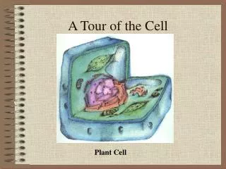
A Tour of the CellThe Organelles Chapter 4 Part II
Reference measurements along the left side marks a 10-fold decrease in size • Most cells are between 1 and 100um in diameter Fig 4.3
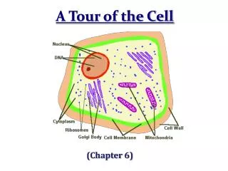
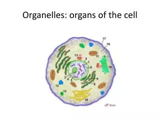
The Nucleus and Ribosomes:Genetic Control of the Cell The cell as a factory: • Nucleus – executive boardroom • Genes – top managers • Proteins - workers
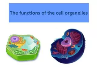
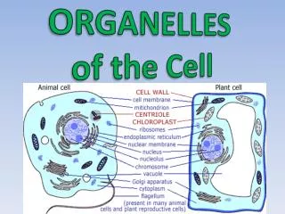
Structure and Function of the Nucleus • Nuclear envelope – double membrane • Nuclear pores – allow selective passage of molecules • Chromatin – DNA molecule and associated proteins • Nucleolus – in association with chromatin - a ball-like mass of fibers and granules produce the component parts of ribosome
Ribosomes • Components are assembled in the nucleus - move through the nuclear pores into the cytoplasm - where they do their work - ribosomes are responsible for protein synthesis • Some ribosomes are suspended in the cytosol - will produce proteins that will remain in the cytosol • Some ribosomes are attached to the outside of the endoplasmic reticulum (ER) - will be incorporated into the cellular membranes or secreted out of the cell
How DNA Controls the Cell • DNA transcribes its coded information into RNA - information in the messenger RNA is used to make proteins - mRNA molecule carries the order to ‘build this specific type of protein’ from the nucleus to the cytoplasm • mRNA exits through the nuclear pores - travels to the cytoplasm where it binds to ribosomes • As a ribosome moves along the mRNA, the genetic message is translated into a protein - a specific amino acid sequence
The Endomembrane System: Cellular Product Manufacturing and Distribution • A eukaryotic cell is partitioned by membranes • Many of the membranous organelles belong to the endomembrane system - includes the endoplasmic reticulum (ER), the Golgi apparatus, lysosomes, and vacuoles
The Endoplasmic Reticulum (ER) • One of the main manufacturing facilities within a cell - produces an enormous variety of molecules • ER is a membranous labyrinth of tubes and sacs running throughout the cytoplasm - membrane separates the internal compartment from the surrounding cytosol • There are 2 distinct types of ER: smooth and rough ER
ER • Flattened sacs of RER and tubes of SER are continuous • ER is continuous with the nuclear envelope Figure 4.10
Rough ER (RER) • Ribosomes are the ‘roughness’ of the RER • Ribosomes of the RER produce 2 main types of proteins: membrane and secretory • Membrane proteins - some are used in the RER • Secretory proteins are exported (secreted) to the fluid outside the cell (extracellular fluid) - cells that secrete a lot of protein are rich in RER • Some poducts are dispatched to other locations in the cell via transport vesicles - membranous spheres that bud off from the ER
Smooth ER (SER) • Smooth ER lacks the surface ribosomes of RER - diversity of enzymes in the SER membrane enables this organelle to perform many functions • Produces lipids, including steroids - cells in ovaries or testes produce sex hormones • SER functions in the detoxification of drugs and other toxins that might be in the bloodstream - enzymes detoxify sedatives, stimulants, some antibiotics - amounts of SER and its enzymes increase after exposure - desensitization or increase tolerance to some drugs can occur and can increase tolerance to other drugs
The Golgi Apparatus (GA) • Named for its discoverer the scientist Camillo Gogi: The GA is a refinery, warehouse, & shipping center - works in close partnership with the ER - receives, refines, stores, distributes the chemical products of the cell - products made in the ER are packaged into transport vesicles to the Golgi where many are modified • GA has 2 sides: – the ‘receiving’ side serves as a dock for the transport vesicles - finished products can be dispatched via the ‘shipping’ side in transport vesicles to other organelles or to the PM
The Golgi Apparatus (GA) • This component of the endomembrane system consists of flattened sacs arranged like a stack of pita bread • A cell may contain a few or hundreds Fig 4.12
Lysosomes (“Breakdown Body”) • A lysosome is a membrane-enclosed sac of digestive enzymes - breaks down macromolecules - compartmentalized for safe digestion without committing ‘suicide’ • Lysosomes have several types of digestive functions: - fuse with tiny cytoplasmic sacs called food vacuoles that expose the food to digestive enzymes - result in small molecules that leave the lysosome to nourish the cell
Digestion of Nutrients Figure 4.13a
Lysosomal Functions • Lysosomes help destroy harmful bacteria - white blood cells ingest bacteria into vacuoles, lysosomal enzymes emptied into these vacuoles rupture the bacterial cell wall • Lysosomes serve as recycling centers for damaged organelles - engulf and digest parts of another organelle to make its molecules available for new organelle construction
Recycling of a Damaged Organelle Figure 4.13b
Embryonic Development • Lysosomes have sculpturing functions - in the embryo lysosomal enzymes destroy cells of the webbing that joins early embryo fingers - lysosomes act as ‘suicide packs’ breaking open to cause programmed cell death (apoptosis)
Lysosomal Storage Diseases • Hereditary disorders in which a person: - lacks one or more digestive enzymes - lysosome become engorged with indigestible substances - interferes with other cellular functions - most are fatal in early childhood • Pompe’s disease, weakening of muscle cells - lack of an enzyme to digest glycogen, accumulation of glycogen in muscle cells, particularly in the heart • Tay-Sachs disease, ravages the nervous system - lack of a lipid-digesting enzyme, accumulation of excess lipids destroy nerve cells
Vacuoles • Membranous sacs that bud from the ER, Golgi, PM - come in different sizes and have a variety of functions • Contractile vacuoles in freshwater protists function as pumps to expel excess water • Central vacuoles of plants – versatile compartment - in seeds (beans, peas) proteins are stockpiled in vacuoles - some absorb water causing plant growth by expansion - in flower petals some may contain pigments that attract pollinating insects - may also contain poisons for protection against plant-eating animals
The first eukaryotic cells are thought to have been protists. They gave rise to fungi, plants, and animals The central vacuole is often the largest organelle in a mature plant cell Fig 4.14
Review of the Endomembrane System • All the organelles of the endomembrane system are related • A product made in one part of this system can eventually exit the cell or become part of another organelle without ever crossing a membrane • Membranes originally made by the ER can eventually become part of the PM through fusion of secretory vesicles
EndomembraneSystem 1) Digestive enzymes made by the RER are transported via vesicles to the GA 2) Lysosomes with processed digestive enzymes bud off from the GA 3) Cell products end up in vacuoles for storage 4) Cell products are secreted from the cell Figure 4.15
Chloroplasts and Mitochondria:Energy Conversion • Cells require a continuous energy supply to do all the work of life • The 2 organelles that produce cellular energy are chloroplasts and mitochondria
Chloroplasts • Most of the living world runs on energy provided by photosynthesis - conversion of light energy from the sun to chemical energy of sugar and other organic molecules • Chloroplasts, organelles in photosynthetic cells of plants and protists perform photosynthesis • Partitioned into 3 major compartments by internal membranes: 1) the space between the 2 membranes that surround the chloroplast 2) the stroma or thick fluid within the chloroplast 3) suspended in that fluid, a network of membrane-enclosed tubes and disks
The Chloroplast: Site of Photosynthesis • Grana - the interconnected stacks of disks • The chloroplast’s solar power packs - structures that trap light energy and convert it to chemical energy Fig 4.16
Mitochondria • Mitochondria are sites of cellular respiration - harvests energy from sugars and other food molecules - converts it to a form of chemical energy called ATP • ATP molecules are a direct energy source for most cellular work • Mitochondria are found in almost all eukaryotic cells • Mitochondria and chloroplasts contain their own DNA that encodes some of their proteins - existence of separate ‘mini-genomes’ is thought to be evidence that these organelles evolved from free-living prokaryotes in the distant past
Mitochondria Structure • An envelope of 2 membranes encloses the mitochondrion, which contains a thick fluid called the matrix • The inner membrane has numerous infoldings called cristae - many of the enzymes and molecules involved in cellular respiration are built into the inner membrane - cristae increase surface area to maximize ATP output
Mitochondrial DNA • Small genomes of mitochondria can affect human health • In 2004, researchers discovered a mutation within one mitochondrial gene - this mutation can cause a condition called metabolic syndrome - can lead to significant health problems, including hypertension and diabetes
The Cytoskeleton: Cell Shape and Movement • A cell’s infrastructure consisting of a network of fibers extending throughout the cytoplasm • The cytoskeleton functions in both support and movement
Maintaining Cell Shape • Provides mechanical support to the cell and maintains its shape - important for cells that lack rigid cell walls • Contains several types of fibers made from different types of proteins: - most important are microtubules, straight, hollow tubes composed of globular proteins called tubulins - filaments are thinner and solid fibers • Provides anchorage and reinforcement for many organelles in a cell - organelles move along tracks made from microtubules - during mitosis microtubules guide the movement of chromosomes
Movement The cell’s cytoskeleton is very dynamic • Can quickly dismantle in one part of the cell by removing protein subunits • Re-form in a new location by reattaching the subunits • Rearrangement of microtubules can provide: - rigidity in a new location - change the cell shape - cause the whole cell or some of its parts to move as in the amoeboid (crawling) motions of the protist Amoeba and some our white blood cells
Rapid tearing down of microtubule subunits and rebuilding of new microtubules is responsible for the crawling movement of organisms like the protist Amoeba Figure 4.18b
Cilia and Flagella • Cilia and flagella are motile appendages or extensions from a cell that aid in locomotion - a specialized arrangement of microtubules functions in the beating of flagella and cilia • Eukaryotic flagella propel the cell by an undulating whiplike motion - occur singularly as in the sperm cells of humans • Cilia are shorter and more numerous - promote movement by a coordinated back-and-forth motion - some extend from nonmoving cells, part of a tissue layer that function to move fluid over the tissue surface
A flagellum usually undulates with a snakelike motion driving a cell as this sperm cell through its fluid environment Cilia are shorter, more numerous, move with a back-and-forth motion. Beating cilia covers this Paramecium, darts rapidly through its watery home Fig 4.19a, b
The cilia lining your respiratory tract sweep mucus with trapped debris out of your lungs Figure 4.19c
The Origin of Membranes • The PM is the boundary of all living cells - logical to hypothesize that they formed early in the evolution of life on Earth • A membrane regulates flow of material across it - can enclose a solution different in composition from the surrounding solution - permits uptake of nutrients and elimination of waste - phospholipids, the key ingredient were probably among the organic molecules that formed from chemical reactions on early Earth and as demonstrated in test tubes phospholipids can spontaneously self-assemble into simple membranes - origin of membranes was likely an early step in evolution of the first cells
Spontaneous Formation of Membranes: A Key Step in the Origin of Life • Phospholipid molecules mixed with water in a test tube, will spontaneously congregate and form water-filled bubbles - primitive membranes • With some ability to control traffic of substances into & out of the sphere Fig 4.20