Download
1 / 1
10 likes | 132 Vues
How can we study pesticides impacts on marine phytoplankton ?. Sabine Stachowski 1 , Harold Anseaume 1 , Dorothée Hureau 2 , Gaël Durand 2 , Denis de la Broise 1 1 LUMAQ, Université de Bretagne Occidentale, 6 rue de l’université, 29334 Quimper, France. Sabine.stachowski@univ-brest.fr

E N D
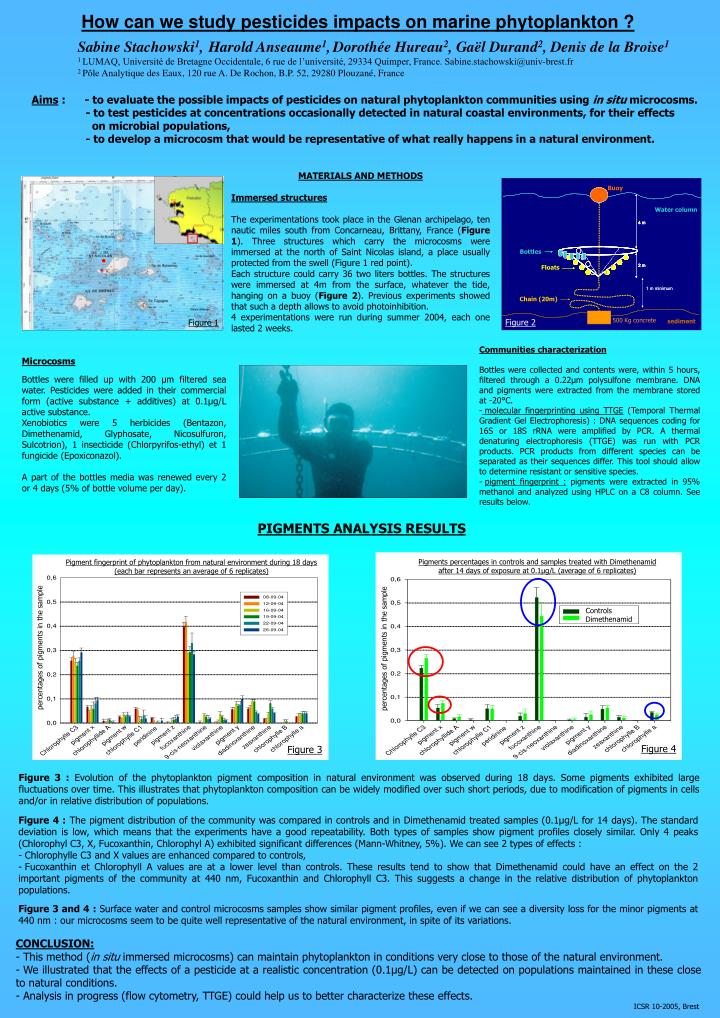
How can we study pesticides impacts on marine phytoplankton ? Sabine Stachowski1,Harold Anseaume1,Dorothée Hureau2, Gaël Durand2, Denis de la Broise1 1 LUMAQ, Université de Bretagne Occidentale, 6 rue de l’université, 29334 Quimper, France. Sabine.stachowski@univ-brest.fr 2 Pôle Analytique des Eaux, 120 rue A. De Rochon, B.P. 52, 29280 Plouzané, France Aims : - to evaluate the possible impacts of pesticides on natural phytoplankton communities using in situ microcosms. - to test pesticides at concentrations occasionally detected in natural coastal environments, for their effects on microbial populations, - to develop a microcosm that would be representative of what really happens in a natural environment. MATERIALS AND METHODS Immersed structures The experimentations took place in the Glenan archipelago, ten nautic miles south from Concarneau, Brittany, France (Figure 1). Three structures which carry the microcosms were immersed at the north of Saint Nicolas island, a place usually protected from the swell (Figure 1 red point). Each structure could carry 36 two liters bottles. The structures were immersed at 4m from the surface, whatever the tide, hanging on a buoy (Figure 2). Previous experiments showed that such a depth allows to avoid photoinhibition. 4 experimentations were run during summer 2004, each one lasted 2 weeks. Buoy Water column Bottles Floats Chain (20m) 500 Kg concrete Figure 2 Figure 1 sediment • Communities characterization • Bottles were collected and contents were, within 5 hours, filtered through a 0.22µm polysulfone membrane. DNA and pigments were extracted from the membrane stored at -20°C. • molecular fingerprinting using TTGE (Temporal Thermal Gradient Gel Electrophoresis) : DNA sequences coding for 16S or 18S rRNA were amplified by PCR. A thermal denaturing electrophoresis (TTGE) was run with PCR products. PCR products from different species can be separated as their sequences differ. This tool should allow to determine resistant or sensitive species. • pigment fingerprint : pigments were extracted in 95% methanol and analyzed using HPLC on a C8 column. See results below. Microcosms Bottles were filled up with 200 µm filtered sea water. Pesticides were added in their commercial form (active substance + additives) at 0.1µg/L active substance. Xenobiotics were 5 herbicides (Bentazon, Dimethenamid, Glyphosate, Nicosulfuron, Sulcotrion), 1 insecticide (Chlorpyrifos-ethyl) et 1 fungicide (Epoxiconazol). A part of the bottles media was renewed every 2 or 4 days (5% of bottle volume per day). PIGMENTS ANALYSIS RESULTS Pigments percentages in controls and samples treated with Dimethenamid after 14 days of exposure at 0.1µg/L (average of 6 replicates) Pigment fingerprint of phytoplankton from natural environment during 18 days (each bar represents an average of 6 replicates) Controls Dimethenamid percentages of pigments in the sample percentages of pigments in the sample Figure 4 Figure 3 Figure 3 : Evolution of the phytoplankton pigment composition in natural environment was observed during 18 days. Some pigments exhibited large fluctuations over time. This illustrates that phytoplankton composition can be widely modified over such short periods, due to modification of pigments in cells and/or in relative distribution of populations. • Figure 4 : The pigment distribution of the community was compared in controls and in Dimethenamid treated samples (0.1µg/L for 14 days). The standard deviation is low, which means that the experiments have a good repeatability. Both types of samples show pigment profiles closely similar. Only 4 peaks (Chlorophyl C3, X, Fucoxanthin, Chlorophyl A) exhibited significant differences (Mann-Whitney, 5%). We can see 2 types of effects : • - Chlorophylle C3 and X values are enhanced compared to controls, • Fucoxanthin et Chlorophyll A values are at a lower level than controls. These results tend to show that Dimethenamid could have an effect on the 2 important pigments of the community at 440 nm, Fucoxanthin and Chlorophyll C3. This suggests a change in the relative distribution of phytoplankton populations. • Figure 3 and 4: Surface water and control microcosms samples show similar pigment profiles, even if we can see a diversity loss for the minor pigments at 440 nm : our microcosms seem to be quite well representative of the natural environment, in spite of its variations. • CONCLUSION: • This method (in situ immersed microcosms) can maintain phytoplankton in conditions very close to those of the natural environment. • We illustrated that the effects of a pesticide at a realistic concentration (0.1µg/L) can be detected on populations maintained in these close to natural conditions. • Analysis in progress (flow cytometry, TTGE) could help us to better characterize these effects. ICSR 10-2005, Brest