Download
1 / 65
660 likes | 909 Vues
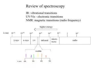
Review of spectroscopy IR: vibrational transitions UV-Vis - electronic transitions NMR: magnetic transitions (radio frequency). N uclear M agnetic R esonance spectroscopy. Some atoms act as tiny magnets. if placed in a magnetic field, will align with or against.

E N D
Review of spectroscopy IR: vibrational transitions UV-Vis - electronic transitions NMR: magnetic transitions (radio frequency)
Nuclear Magnetic Resonance spectroscopy Some atoms act as tiny magnets if placed in a magnetic field, will align with or against -1/2 spin state is slightly higher E: Slightly > 50% of atoms are in +1/2 state 5.1A
Angular frequency of precession: w Values of w are in radio frequency range varies with strength of B0! 5.1A
The resonance condition: If sample is hit with radiation of frequency n = w, we have resonance condition: spin flip! That frequency of radiation is absorbed.
Protons in different chemical environments have different resonance frequencies! Key concept: chemical equivalence/nonequivalence
These molecules have several different ‘sets’ of protons 5.2
Enantiotopic/homotopic protons have equivalent resonance frequencies 5.2
Practice: How many sets of protons? (ie. how many 1H-NMR signals?
The 1H-NMR experiment • Put sample in strong external magnetic field • Ha protons precess at wa, Hb protons at wb • Hit sample with radiation in Rf range of frequencies • Ha protons absorb radiation at wa, undergo ‘spin flip’ • Hb protons absorb radiation at wb, undergo ‘spin flip’ • Detector records which frequencies were absorbed, and intensity of each absorbance 5.3A
We must use solvents without protons! (Used to use CCl4 commonly, but it’s carcinogenic) 5.3A
The chemical shift (refer to 1H-NMR spectrum of methyl acetate) TMS (tetramethyl silane) is used as a standard: set the resonance frequencies of these 12 equivalent protons equal to zero • Record the resonance frequencies of the protons in your sample relative to TMS protons, expressed as ppm • eg. 7.1 T magnetic field, TMS protons resonate at 300,000,000 Hz (300 MHz) • Ha protons resonate at 300,000,621 Hz, which is 2.07 ppm higher than TMS protons • Hb protons resonate at 300,001,104 Hz, which is 3.68 ppm higher than TMS protons
Remember: Resonance frequencies vary with strength of B0! but . . . when expressed in terms of ppm relative to TMS, the number does not change - this is why we use ‘chemical shift’ rather than Hz (or wavelength) example: A proton has a chemical shift of 4.50 ppm. a)What is its resonance frequency, expressed in Hz, in a 300 MHz instrument (ie an instrument with a 7.1 Tesla magnet, where TMS protons resonate at 300 MHz)? b)What is its chemical shift expressed in Hz? c) What is its resonance frequency in a 100 MHz instrument?
Chemical shift is abbreviated by d Higher chemical shifts are said to be ‘downfield’ Most protons in organic compounds have chemical shifts from 0-12 ppm relative to TMS TMS is (usually) no longer added to sample: resonance frequency of deuterium in solvent is used as the actual reference point (but 0 ppm is still defined as TMS signal)
Signal integration The area under a 1H-NMR signal (integrations) corresponds to how many protons cause the signal In methyl acetate, the Ha signal and Hb signal both represent three protons. Thus, the area under these signals is (approx) equal In p-xylene, Ha corresponds to six protons, Hb to four. Ratio of peak integrations is 6 to 4, or 1.5 to 1
The basis for magnetic non-equivalence (why do different protons have different chemical shifts?) The shielding effect: nearby electrons create small magnetic fields in opposition to B0. These are called ‘induced fields’. 5.4A
The deshielding effect Electronegative atoms pull electrons away from nearby protons Protons are ‘deshielded’, experience stronger Beff Stronger Beff means higher resonance frequency: higher (downfield) chemical shift 5.4A
Diamagnetic anisotropy isotropy = ‘sameness’ anisotropy = ‘difference’ Why is benzene chemical shift so far downfield? . . . more than just normal deshielding! 5.4B
Field from a magnet is anisotropic at point A, you sense a field pushing north at point B, you sense a field pushing south 5.4B
6 aromatic electrons form ring current, opposed to B0 But for benzylic protons, the ring current field is aligned with B0 - makes Beff stronger! Strong deshielding effect. 5.4B
(exercise 5.5) Extreme case: outer protons are 8.9 ppm inner protons are -1.8 ppm (upfield of TMS signal!) inside the ring, aromatic ring current is strongly shielding
Hydrogen-bonding protons (amines, alcohols, phenols) have variable variable chemical shifts, often >4 ppm. H-bonding patterns effect chemical shift Often slightly broad peaks (see spectrum next slide)
Spin-spin coupling Ha 5.5A
Ha signal in 1,1,2-trichloroethane is a doublet ‘split’ by Hb 5.5A
Hb signal is a triplet ‘split’ by Ha Ha and Hb are coupled - their spins interact 5.5A
Splitting is seen between protons that are separated by three bonds or less n neighbors leads to n + 1 sub-peaks H-bonded protons generally do not show coupling
ethyl acetate 5.5A
Coupling constants J is expressed in Hz, not ppm - does not depend on strength of B0! Notice: same value of J! Function of interaction between Ha and Hb 5.5B
Complex coupling methyl acrylate 5.5C
(exercise 5.9 asks you to construct a splitting diagram for Hb) 5.5C
Often the ‘n+1 rule’ holds even when protons are non-equivalent . . . if J values are close 5.5C
Sometimes there is too much overlapping: just call it a multiplet (m) 5.5C
13C-NMR Spectroscopy • Differences from 1H-NMR: • Only ~ 1% of carbons are 13C - much weaker signal • Integration not meaningful: signal intensities vary (eg. carbonyl carbon signals are very weak) • Where TMS protons resonate at 300 MHz, 13C resonates at 75 MHz • Chemical shift range is wider - more than 200 ppm
Broadband decoupling: turns off C-H splitting, so we see only singlets 5.6A