Download
1 / 41
420 likes | 659 Vues
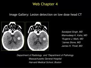
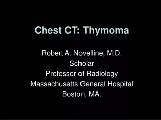
Web Chapter 3. Image Gallery: Lesion detection on low dose chest CT. Sarabjeet Singh, MD Mannudeep K. Kalra, MD *Eugene J. Mark, MD *James Stone, MD James H. Thrall, MD. Department of Radiology and *Department of Pathology Massachusetts General Hospital Harvard Medical School, Boston.

E N D
Web Chapter 3 Image Gallery: Lesion detection on low dose chest CT Sarabjeet Singh, MD Mannudeep K. Kalra, MD *Eugene J. Mark, MD *James Stone, MD James H. Thrall, MD Department of Radiology and *Department of Pathology Massachusetts General Hospital Harvard Medical School, Boston
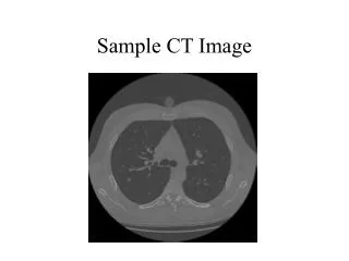
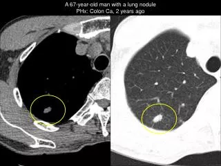
Do you see any abnormal findings in this transverse CT image?
Do you see a nodule in right lower lobe, paraseptal emphysema, and centrilobular emphysema?
Do you see any additional finding in higher dose images ? Image A Image B Image C nodule in right lower lobe, paraseptal emphysema in the left lung, centrilobular emphysema
Do you see any abnormal findings in this transverse CT image?
Do you see.. Solid and cystic lesion in left lower lobe abutting the major fissure, ground glass opacity lateral to descending aorta, dependent atelectasis bilaterally?
Do you see any additional finding in higher dose images ? Image A Image B Image C Solid and cystic mass lesion in LLL, ground glass opacity lateral to descending aorta, and dependent atelectasis bilaterally
Do you see any abnormal findings in this transverse CT image?
Do you see multiple peribronchovascular and subpleural nodules consistent with pulmonary sarcoidosis?
Do you see any additional finding in higher dose images ? Image A Image B Image C Multiple peribronchovascular and supleural nodules consistent with pulmonary sarcoidosis
Do you see any abnormal findings in this transverse CT image?
Do you see subpleural opacity in the anterior left upper lobe, right major fissure nodule, and thickened left major fissure?
Do you see any additional finding in higher dose images ? Image A Image B Image C Subpleural opacity in the anterior LUL, right fissural nodule, thickened left major fissure
Do you see any abnormal findings in this transverse CT image?
Do you see paraseptal and bullous emphysema, and nodule in the right upper lobe?
Do you see any additional finding in higher dose images ? Image A Image B Image C Paraseptal and bullous emphysema. Nodule in the right upper lobe.
Do you see any abnormal findings in this transverse CT image?
Do you see patchy air space opacity in the right lower lobe, Multiple right fissural nodules, and right middle lobe non-calcified nodule?
Do you see any additional finding in higher dose images ? 540 mA; 20 mGy 270 mA; 10 mGy 135 mA; 5.0 mGy Patchy air space opacity in the RLL Right fissural nodules RML nodule 72 mA; 2.7 mGy 36 mA; 1.3 mGy
Do you see any abnormal findings in this transverse CT image?
Do you see branching nodular opacities in right middle lobe, patchy consolidation in bilateral lower lobes (right greater than left) and right fissural effusion?
Do you see any additional finding in higher dose images ? 270 mA; 10 mGy 135 mA; 5.0 mGy 540 mA; 20 mGy Branching nodular opacities in RML, patchy consolidation in RLL right fissural effusion seen at all dose levels 72 mA; 2.7 mGy 36 mA; 1.3 mGy
Do you see any abnormal findings in this transverse CT image?
Do you see mediastinal lymph nodes, and calcified plaques in the ascending and descending thoracic aorta?
Do you see any additional finding in higher dose images ? Image A Image B Image C Mediastinal lymph node, aortic calcification in ascending and descending thoracic aorta
Do you see any abnormal findings in this transverse CT image?
Do you see left pleural effusion and left fissural effusion.
Do you see any additional findings on higher dose images Image A Image B Image C Left pleural effusion and left fissural effusion.
Do you see any abnormal findings in this transverse CT image?
Do you see calcified and non-calcified mediastinal lymph nodes?
Do you see any additional findings on higher dose images? Image A Image B Image C Calcified and non-calcified mediastinal lymph nodes
Do you see any abnormal findings in this transverse CT image?
Do you see left ventricular apical calcification suggestive of old myocardial infarction in the LAD territory ?
Do you see any additional findings on higher dose images? Left ventricular apical calcification suggestive of old myocardial infarction in the LAD territory
Do you see any abnormal findings in this transverse CT image?
Do you see bilateral pleural effusions with bilateral lower lobe atelectasis, enlarged right atrium and ventricle, and pericardial effusion?
135 mAs; 10 mGy 72 mAs; 5.0 mGy 40 mAs; 2.7 mGy 20 mAs; 1.3 mGy B/L pleural effusions with atelectasis, enlarged right atrium and ventricle, pericardial effusion Distinction between pleural effusion and atelectasis is not clear on 20 mAs image.
Do you see lytic lesion with sclerotic rim in the head of the left humeral head?
Do you see any additional findings on higher dose images? 135 mAs; 10 mGy 72 mAs; 5.0 mGy 40 mAs; 2.7 mGy 20 mAs; 1.3 mGy
Thank You Please contact for any questions Sarabjeet Singh, MD: ssingh6@partners.org Mannudeep K Kalra, MD: mkalra@partners.org