Cellular Aspects of Central Nervous System Pathology
Explore cellular markers of injury in the brain, including red neurons, axonal injury, and cerebral edema. Study the pathology of brain tumors and neurodegenerative diseases. Gain insights into CNS disorders such as meningitis, hydrocephalus, and multiple sclerosis. Recommended textbook: Robbins Basic Pathology, 9th Edition. Make sure to grasp key principles from the lecture contents.

Cellular Aspects of Central Nervous System Pathology
E N D
Presentation Transcript
Course contents • Cellular aspects of nervous system injury • Pathology of brain tumors • Multiple sclerosis • Cerebrovascular accidents • Neurodegenerative brain disease • Meningitis • Congenital malformations and hydrocephalus Please make sure you got your copy from the course description by the end of this lecture
CNS pathology course • Recommended textbook: • Vinay Kumar, Abul K. Abbas, Nelson Fausto, & Richard Mitchell , Robbins Basic Pathology, 9th Edition. • The course guidelines including the lectures contents are very important. Make sure to read them carefully. • You are requested to study all the Key principles, even if they were not discussed in the lectures.
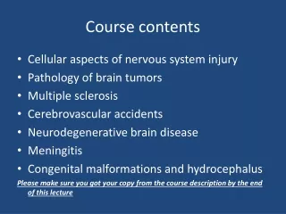
Red neurons (A) Spheroids (B) Chromatolysis (C)
Markers of Neuronal Injury • Red neuron: • Within 12 hours of an irreversible hypoxic/ischemic insult, acute neuronal injury becomes evident even on routine hematoxylin and eosin (H & E) staining: • shrinkage of the cell body • pyknosis of the nucleus • disappearance of the nucleolus • loss of Nissl substance • intense eosinophilia of the cytoplasm ("red neurons“) • Acute injuries typically result in breakdown of the blood-brain barrier and variable degrees of cerebral edema
Markers of Neuronal Injury • intracellular inclusions: • Nuclear or cytoplasmic aggregates of stainable substances, usually proteins. • Example: Negri bodies in rabies
Markers of Neuronal Injury • dystrophic neurites: • A neurite refers to any projection from the cell body of a neuron • In some neurodegenerative diseases, neuronal processes become thickened and tortuous; these are termed dystrophic neurites
Markers of Neuronal Injury • Axonal injury • Injured axons undergo swelling (called spheroids) and show disruption of axonal transport • Evidence of injury can be highlighted by silver staining or immunohistochemistry for axonally transported proteins such as amyloid precursor protein • Axonal injury also leads to cell body enlargement and rounding, peripheral displacement of the nucleus, enlargement of the nucleolus, and dispersion of Nissl substance (from the center of the cell to the periphery, so-called central chromatolysis)
Immunostains with antibodies to Beta Amyloid Precursor Protein (BAPP) can detect the axonal lesions in 2-3 hours after the injury (diffuse axonal injury)
Markers of Neuronal Injury • Diffuse axonal injury • As many as 50% of patients who develop coma shortly after trauma, even without cerebral contusions, are believed to have white matter damage and diffuse axonal injury • Widespread injury to axons within the brain can be very devastating • The movement of one region of brain relative to another is thought to lead to the disruption of axonal integrity and function • Diffuse axonal injury is characterized by the wide but often asymmetric distribution of axonal swellings that appears within hours of the injury and may persist for much longer • These are best demonstrated with silver stains or by immunohistochemistry for proteins within axons
Cerebral Edema • The accumulation of excess fluid within the brain parenchyma • Two types, which often occur together particularly after generalized injury: • Vasogenic edema: • The integrity of the normal blood-brain barrier is disrupted, allowing fluid to shift from the vascular compartment into the extracellular spaces of the brain • can be either localized (e.g., increased vascular permeability due to inflammation or in tumors) or generalized
Cerebral Edema • Cytotoxic edema: • An increase in intracellular fluid secondary to neuronal and glial cell membrane injury, as might follow generalized hypoxic-ischemic insult or after exposure to some toxins
Astrocytes in Injury and Repair • Astrocytes are the principal cells responsible for repair and scar formation in the brain, a process termed gliosis • In response to injury: • Astrocytes undergo both hypertrophy and hyperplasia • The nucleus enlarges and becomes vesicular, and the nucleolus is prominent • The previously scant cytoplasm expands to a bright pink, somewhat irregular swath around an eccentric nucleus, from which emerge numerous stout, ramifying processes (gemistocyticastrocyte) • In settings of long-standing gliosis, astrocytes have less distinct cytoplasm and appear more fibrillar(fibrillaryastrocytes)
Astrocytes in Injury and Repair • There is minimal extracellular matrix deposition: Unlike the repair after injury elsewhere in the body, fibroblasts participate in healing after brain injury only to a limited extent (usually after penetrating brain trauma or around abscesses)
Astrocytes in Injury and Repair • Rosenthal fibers are thick, elongated, brightly eosinophilic protein aggregates that can be found in astrocytic processes in chronic gliosis and in some low-grade gliomas Which tumor exhibits Rosenthal fibers?
Oligodendrocytes in Injury and Repair • Produce myelin • Exhibit a limited spectrum of specific morphologic changes in response to various injuries • In progressive multifocal eukoencephalopathy, viral inclusions can be seen in oligodendrocytes, with a smudgy, homogeneous-appearing enlarged nucleus
Ependymal cells in Injury and Repair • line the ventricular system and the central canal of the spinal cord • Certain pathogens, particularly cytomegalovirus (CMV), can produce extensive ependymal injury, with typical viral inclusions
Microglial nodule Neuronophagia
Microglia in Injury and Repair • Microglia: • Bone marrow-derived cells • Function as the phagocytes of the CNS • When activated, they proliferate and become more evident • They may be recognizable as activated macrophages in areas of: • Demyelination • Organizing infarct • Hemorrhage • They develop elongated nuclei (rod cells) in neurosyphilis or other infections • When these elongated microglia form aggregates at sites of tissue injury, they are termed microglial nodules • Similar collections can be found congregating around portions of dying neurons, termed neuronophagia (e.g. viral encephalitis).
Markers of peripheral nerve injury • Most peripheral neuropathies can be subclassified as either axonal or demyelinating, even though some diseases exhibit mixed features
Markers of peripheral nerve injury • Axonal neuropathies: • Caused by insults that directly injure the axon • The entire distal portion of an affected axon degenerates • Axonal degeneration is associated with secondary myelin loss a process sometimes referred to as Wallerian degeneration • Regeneration takes place through axonal regrowth and subsequent remyelination of the distal axon • The morphologic hallmark of axonal neuropathies is a decrease in the density of axons, which in electrophysiologic studies correlates with a decrease in the strength of amplitude of nerve impulses.
Markers of peripheral nerve injury • Segmental demyelination: • Demyelinating neuropathies are characterized by damage to Schwann cells or myelin with relative axonal sparing, resulting in abnormally slow nerve conduction velocities • Demyelination typically occurs in individual myelin internodes randomly; this process is termed segmental demyelination • Morphologically, demyelinating neuropathies show a relatively normal density of axons and features of segmental demyelination and repair >> recognized by the presence of axons with abnormally thin myelin sheaths and short internodes
Homework • Define Corpora amylacea. Where and when they are deposited in the CNS? Note: Source: your choice!