Download
1 / 87
980 likes | 1.56k Vues
Learn about ECG findings in pacemaker malfunction, basic principles of pacing, rhythm recognition, dual-chamber systems, and more. Understand sensitivity, undersensing, oversensing, and magnet operation in pacemakers.

E N D
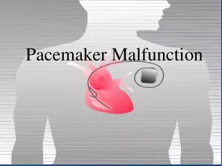
ECG in Pacemaker Malfunction Sriram Rajagopal, Southern Railway Hospital, Perambur,Chennai
ECG in “Pacemaker Malfunction”(ECGs in Pacemaker Function Assessment) Sriram Rajagopal, Southern Railway Hospital, Perambur,Chennai
Implantable Pacemaker Systems Contain the Following Components: Lead wire(s) Implantable pulse generator (IPG)
Pseudomalfunctions Pseudomalfunctions are defined as: • Unusual • Unexpected • Eccentric ECG findings that appear to result from pacemaker malfunction but that represent normal pacemaker function
ECGs in Patients with Pacemakers Overview : • Basic principles of pacing • Background information required • Systematic approach • Examples with single chamber pacing • Examples with dual chamber pacing
ECGs in Patients with Pacemakers Overview : • Basic principles of pacing • Background information required • Systematic approach • Examples with single chamber pacing • Examples with dual chamber pacing
ECGs in Patients with Pacemakers What do pacemakers do ? • Pace • Sense – ( what ? ) • Respond – Inhibit , Trigger or Dual • Respond to increased metabolic demand by providing rate responsive pacing • Provide diagnostic information stored by the pacemaker
Timing Intervals Are Expressed in Milliseconds • One millisecond = 1 / 1,000 of a second
Rate Conversion • Conversion • Pacing rate in PPM divided by 60,000 = ms • 60,000 / 60 PPM = 1000 ms • Interval in ms divided by 60,000 = PPM • 60,000 / 1000 ms = 60 PPM 60,000 ms BPM
Paced Rhythm Recognition VVI / 60
Paced Rhythm Recognition VVI / 60
Paced Rhythm Recognition AAI / 60
Noncapture is Exhibited By: • No evidence of depolarization after pacing artifact Loss of capture
No Output • Pacemaker artifacts do not appear on the ECG; rate is less than the lower rate Pacing output delivered; no evidence of pacing spike is seen
Fusion Beat • Definition: The combination of an intrinsic beat and a paced beat. • The morphology varies; in other words, a fusion beat doesn’t really look like a paced beat or an intrinsic beat. • The pacemaker and the patient contribute to depolarization in Fusion beats.
Fusion • Ventricular Fusion
Pseudofusion Beat • Definition: A pacing pulse falls on an intrinsic beat. The pacing pulse is ineffective and the intrinsic complex is not altered. • The pacemaker does NOT contribute to depolarization in Pseudofusion beats.
Pseudofusion • Ventricular Pseudofusion
Dual-Chamber Systems Have Two Leads: • One lead implanted in the atrium • One lead implanted in the ventricle
Paced Rhythm Recognition DDD / 60 / 120
Paced Rhythm Recognition DDD / 60 / 120
Paced Rhythm Recognition DDD / 60 / 120
Paced Rhythm Recognition DDD / 60 / 120
Sensing • Sensing is the ability of the pacemaker to “see” when a natural (intrinsic) depolarization is occurring • Pacemakers sense cardiac depolarization by measuring changes in electrical potential of myocardial cells between the anode and cathode
Sensitivity – The Greater the Number, the Less Sensitive the Device to Intracardiac Events
Sensitivity 5.0 2.5 Amplitude (mV) 1.25 Time
Sensitivity 5.0 2.5 Amplitude (mV) 1.25 Time
Sensitivity 5.0 2.5 Amplitude (mV) 1.25 Time
Undersensing . . . • Pacemaker does not “see” the intrinsic beat, and therefore does not respond appropriately Scheduled pace delivered Intrinsic beat not sensed VVI / 60
VVI / 60 Oversensing • An electrical signal other than the intended P or R wave is detected ...though no activity is present Marker channel shows intrinsic activity...
ECGs in Patients with Pacemakers Overview : • Basic principles of pacing • Background information required • Systematic approach • Examples with single chamber pacing • Examples with dual chamber pacing
ECGs in Patients with Pacemakers Basic Data : • Clinical details – age , indication for pacing, time since implant etc • Type of pacemaker • Programmed parameters • Magnet Behaviour • Special features
Magnet Operation • Magnet application causes asynchronous pacing at a designated “magnet” rate
Rate Responsive Pacing • An accelerating or decelerating rate may be perceived as anomalous pacemaker behavior VVIR / 60 / 120
Hysteresis • Allows a lower rate between sensed events to occur; paced rate is higher Hysteresis Rate 50 ppm Lower Rate 70 ppm
ECGs in Patients with Pacemakers Overview : • Basic principles of pacing • Background information required • Systematic approach • Examples with single chamber pacing • Examples with dual chamber pacing
ECGs in Patients with Pacemakers Single chamber pacing : • Identify underlying intrinsic rhythm if any ( in each chamber) • Verify appropriate sensing ( if possible ) • Verify capture • Measure base rate and check if appropriate • Identify any variations in intervals and interpret • Identify causes of inappropriate sensing or pacing
ECGs in Patients with Pacemakers Overview : • Basic principles of pacing • Background information required • Systematic approach • Examples with single chamber pacing • Examples with dual chamber pacing
Single Chamber ECG Analysis Programmed Parameters Mode………………………………………….. VVI Base Rate……………………………………….. 70 ppm Magnet Response…………………….. Battery Test Hysteresis Rate………………………………… Off ppm T Temporary programmed value 1.0 Second 7 Mar 2000 23:20 1
ECG #1 • VVI • Normal Capture and Sensing 1
Single Chamber ECG Analysis Programmed Parameters Mode………………………………………….. VVI Base Rate……………………………………….. 70 ppm Magnet Response…………………….. Battery Test Hysteresis Rate………………………………… Off ppm T Temporary programmed value 1.0 Second Jun 14 1999 2:57 pm 2
ECG #2 • VVI • Normal Sensing • Capture unknown 2
ECG #3 • VVI • Normal Capture and Sensing with initiation of Hysteresis 3
ECG #4 • VVI • Loss of Ventricular Sensing • Ventricular Undersensing with Functional loss of capture on the second to last beat 4
ECG #5 • VVI • Loss of Ventricular capture with functional loss of sensing 5
ECG #6 • VVI • Normal Capture • Ventricular Oversensing 6