Download
1 / 53
530 likes | 541 Vues
This article explores the reasons why cells divide, including the limited capacity for DNA and nutrient transport across the cell membrane. It also discusses the problems that arise when cells grow too large and the process of cell division to solve these issues. The concepts of mitosis, cytokinesis, and the cell cycle are covered.

E N D
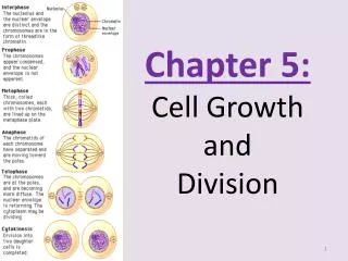
Chapter 10Cell Growth & DivisionSection 10-1 Cell Growth Read 1st Paragraph Fig. 10-1
Limits To Cell Growth • 2 reasons why cells divide rather than get larger. • They don’t have enough DNA. • They can’t move enough nutrients & wastes across the cell membrane.
DNA “Overload” • Remember: • DNA control’s cell function. • DNA is stored in the nucleus. • When a cell is small, the info in DNA is able to meet the cell’s needs. • But, as a cell increases in size, it doesn’t make more DNA. • If it were to keep growing, there would be an “Information Crisis”.
Compare the DNA in the nucleus to that of a small library. • Read P. 241 • Middle paragraph
Exchanging Materials • Recall that food, H2O, & O2pass through CM’s easily by passive & active transport. • Waste also leaves the cell through the cell membrane.
The rate at which these exchanges take place depends on the surface area of the cell. • Surface area =Total area of the cell membrane (CM). • However, the rate at which food & O2are taken in, & wastes produced depends on the cell’s volume. • Volume =How much space is in the cell.
Surface Area –Volume Ratio • Imagine a cell shaped like a cube. • p. 243, fig. 10-2 Follow along! • Important things to note: • Volume increases much faster than surface area. • This causes the surface area-volume ratio to decrease. • This causes serious problems for the cell.
http://plaza.ufl.edu/alallen/pgl/modules/rio/stingarees/module/why.htmlhttp://plaza.ufl.edu/alallen/pgl/modules/rio/stingarees/module/why.html
Surface Area to Volume Ratio Cell Size Surface Area (length x width x 6) Volume (length x width x height) Ratio of Surface Area to Volume
Use the street –traffic analogy. • Read p. 242, bottom paragraph. • If a cell got too big, it would have trouble getting enough O2 & nutrients in, & waste out.
Division of the Cell • Before cells get too big they divide. • Division creates 2“daughter cells”. • This is called cell division. • Before cell division occurs, the cell copies its DNA. • This solves the problem of info shortages. • Cell division also solves the volume problem. • By reducing size, the cell reduces volume.
Section 10-2 Cell Division • Read p. 244 top paragraph • In prokaryotes (pro-k), cell division is just the splitting of the cell into 2 parts. • In eukaryotes(eu-k), cell division is complex & happens in 2 stages: • Mitosis • Cytokinesis
Reproduction through mitosis is considered asexual. • All the cells are identical to the parent cell. • What are the resulting cells called?
Chromosomes • In eukaryotes, genetic info is carried by chromosomes. • Chromosomes are made of DNA & proteins. • Every org. has a certain # of chromosomes: Examples: • Fruit flies =8 • Carrot cells = 18 • Human cells = 46 • Dog = 78 • Sweet potato = 90
Chromosomes are normally visible only during cell division. • The rest of the time they are in a form known as chromatin. Before cell division happens, chromosomes are copied. For this reason, each chromosome consists of TWOidentical“sister”chromatids.See p. 244, Fig. 10-3
When cells divide, the “sister” chromatids separate from each other. • 1 chromatid goes to each of the 2 new cells.
Chromatids are held together by a centromere. • Normally located @ the middle of the chromatid • Some can lie near the ends. • A human cell contains 46 chromosomes (sometimes referred to as 23 pairs) • Each consisting of 2 chromatids.
The Cell Cycle • Cell cycle –series of events that cells go through as they grow & divide. • Interphase –period b/t cell divisions. • During each cell cycle: • A cell grows • Prepares for division • Divides to form 2 daughter cells • p. 245, fig. 10-4
TheCell Cycle G1 Consists of 4 phases: S • G1 Phase • S Phase • G2 Phase • M Phase G2
Events of the Cell Cycle • Most of a cell’s life is spent in interphase. • Interphase is divided into 3 phases: • G1 • Cells do most of their growing. • New proteins & organelles are also made. • S • Chromosomes are copied. • Synthesis of DNA molecules takes place. • G2 • Shortest of the 3 phases. • Organelles & molecules required for cell division are made. When the G2 phase is done, the cell is ready to enter the Mphase. a.k.a. = cell division
Mitosis • Divided into 4 phases: • Prophase • Metaphase • Anaphase • Telophase • Depending on the cell, mitosis can last from minutes to days. • PMAT
Prophase • 1st & longest phase. • Chromosomes become visible. • Centrioles separate & take up positions on opposite sides of the nucleus. Centrioles -2 tiny structures located near the nuclear envelope.
Spindle–a fanlike Microtubule structure that helps separatechromosomes. • Plant cells don’t have centrioles. • Near the end of prophase: • Chromosomes coil more tightly. • The nucleolus disappears. • The nuclear envelope breaks down.
Metaphase • 2nd phase • Often lasts only a few minutes. • Chromosomes line up at the midline or equator of the cell. • The centromeres of each chromosome connect to the spindle.
Anaphase • 3rd phase • During anaphase: • The centromeres joining the sister chromatids, separate & become individual chromosomes. • These chromosomes then move to opposite ends of the cell called poles. • Anaphase ends when the chromosomes stop moving.
Telophase • The 4th & final phase. • Chromosomes begin to disperse into a tangle of dense material. • The nuclear envelope reforms around the chromosomes. • The spindle breaks apart. • The nucleolus becomes • visible again.
M phase (Mitosis) Interphase G1 phase S phase G2 phase Prophase Metaphase Anaphase Telophase Cell Cycle includes is divided into is divided into
Interphase Prophase Metaphase Anaphase Telophase
Cytokinesis • Cytokinesis –the division of the cytoplasm. • Cytokinesis usually occurs at the same time as telophase. • It occurs in primarily 2 ways: • By pinching off b/t the 2 daughter cells equally. • Done in animal cells. • By building a cell plate b/t the 2 cells. • Done in plant cells.
Mitosis Video • http://www.johnkyrk.com/mitosis.html • https://www.youtube.com/watch?v=TlYqh5OfJFk
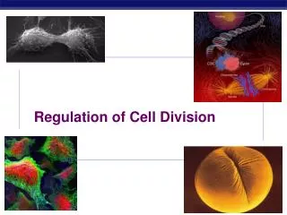
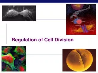
Section 10-3 Regulating the Cell Cycle
Note! • Not all cells move through the cell cycle at same rate. • Ex: • Muscle cells & nerve cells don’t divide at all, once they are developed. • Skin cells, digestive cells, & cells in bone marrow that make blood cells, divide rapidly throughout our life. • Rapidly dividing cells can pass through a cell cycle every few hrs.
Controls of Cell Divisionfig.10-7 • This illustration shows that cell division can be turned on & off. • The same thing happens when we have a cut in the skin or a broken bone. • New cells are made until the wound is healed, then they stop dividing.
Fig. 10-7 • Cells in a petri dish will continue to grow until they come into contact w/ other cells.
Cell Cycle Regulators • In the 1980’s, scientists discovered what controls the cell cycle. • The substance was a protein called cyclin. • Cyclin regulates the cell cycle. • Since this discovery, scientists have found other proteins that are involved in the cell cycle. • They call all these proteins cyclins. p. 251, fig. 10-8
Fig. 10-8 A sample of cytoplasm is removed from a cell in mitosis. The sample is injected into a second cell in G2 of interphase. As a result, the second cell enters mitosis.
There are 2 types of regulatory proteins: Internal Regulators External Regulators
Internal Regulators • Made up of proteins that respond to events inside the cell. • Allows the cell to only proceed when certain events have happened. • Ex: • Cells wont move to the next phase in mitosis until all the right steps have been completed.
External Regulators • Made up of proteins that respond to events outside the cell. • They direct cells to speed up or slow down the cell cycle. • Important during wound healing & embryonic development.
Uncontrolled Cell Division • Cancer –a disorder in which some of the body's own cells lose the ability to control growth. • Cancer cells don’t respond to the signals that regulate the growth of most cells. • As a result they divide uncontrollably & form masses of cells called tumors. • Tumors can damage surrounding tissues. • Cancer cells can also move throughout the body & cause serious problems, or even death.