Download
1 / 79
790 likes | 914 Vues
This educational resource presents an in-depth examination of shoulder syndromes, particularly focusing on the complex anatomy and biomechanics of the shoulder joint. It covers key topics including the anatomy of the shoulder complex, its functional motions, and common shoulder problems. Emphasizing the importance of effective evaluation, it guides practitioners in performing functional and special tests to diagnose shoulder injuries. Recognizing the prevalence of shoulder pain in the active population, this guide serves as a vital tool for sports medicine professionals.

E N D
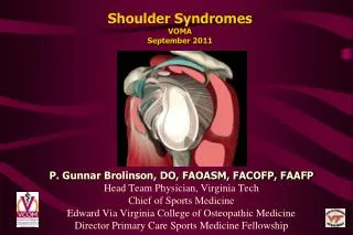
Shoulder SyndromesVOMASeptember 2011 P. Gunnar Brolinson, DO, FAOASM, FACOFP, FAAFP Head Team Physician, Virginia Tech Chief of Sports Medicine Edward Via Virginia College of Osteopathic Medicine Director Primary Care Sports Medicine Fellowship
Objectives • Review anatomy of the shoulder complex • Review motions of the shoulder complex • Describe the functional biomechanical evaluation of the shoulder • Understand and be able to perform an evaluation of shoulder using various functional and special tests • Review some common shoulder problems
Introduction • Shoulder injury is very common in the active patient population. • It is a complex joint and presents unique challenges to diagnosis and subsequent treatment.
Introduction • Shoulder Pain is the third most common MS complaint in primary care offices • Second to knee pain for referrals to ortho/sports medicine physicians • Incidence 25/1000 patients • Peak incidence in 50-70 year olds • 8-13% of athletic injuries involve the shoulder
Introduction • The shoulder complex is a loosely constructed highly mobile complex of bones, muscles and ligaments. • It is designed for increased mobility to the upper extremity with only sufficient stability to provide a proper foundation for muscular function which is vital for the performance of sports or activities of daily living (ADL)
Introduction • Effective diagnosis and treatment of the shoulder requires a mastering of the relationship of structure and function of this complicated joint.
Anatomy • It is composed of 3 joints (sternoclavicular, acromioclavicular and glenohumeral) and one articulation (scapulothoracic). • All four work together in a synchronous rhythm for full range dynamic motion.
Anatomy…SC Joint • The sternoclavicular joint (SCJ) enables the humerus to achieve 180 degrees of Abduction. • It is a saddle shaped joint made up of the medial end of the clavicle, the manubrium sternum and the cartilage of the 1st rib. • There is an articular disc separating the surfaces which adds strength to the joint.
Anatomy…AC Joint • A plane synovial joint that augments the range of motion (ROM) in the humerus. • It is made up of the acromiom process of the scapula and the lateral edge of the clavicle.
Anatomy…AC Joint • It is surrounded by a fibrous capsule and an articular disc separates the surfaces. • Primary strength is supplied by the acromioclavicular and coracoclavicular ligaments • trapezoid ligament • conoid ligaments
AC Joint • Type I 17% • Type II 43% • Type III 40% • Type III found in up to 80% of RC tears • Compared with 3% in Type I
AC Joint/Subacromial Articulation • Impingement • Greater tubercle • Acromion • Coracoacromial ligaments • Supraspinatus tendon • Between 48-72% of shoulder pain in PCP office is subacromial impingement
Anatomy…GH Joint • A multi-axial ball and socket joint surrounded by a capsule. • Most of the support is provided by the rotator cuff muscles.
Anatomy of GH Joint • The glenoid labrum is a ring of fibrocartilage that surrounds and deepens the glenoid fossa which increases the available contact area by approximately 70%.
Functional Anatomy…GH Joint • The relaxed position of the humerus has it placed in the upper portion of the glenoid cavity. • Contraction of the rotator cuff muscles pulls the humerus down into the lower/wider portion of the glenoid cavity. • Without the “dropping down”, full Abduction is impossible.
GH Joint • Static stabilizers • Labrum • Capsule • Adhesion-cohesion • Intra-articular pressure • Dynamic stabilizers • RC muscles • Deltoid • Long head of biceps • Scapulothoracic muscles • Proprioceptive feedback
GH Joint…Static Restraints • Labrum • Ring of fibrocartilage • Deepens the glenoid fossa • Increases contact area ~70% • Ligaments • Superior Glenohumeral • Middle Glenohumeral • Inferior Glenohumeral (important when shoulder is abducted and externally rotated)
The Scapulothoracic Articulation • The scapula serves as a mobile platform from which the upper limb operates. • It is made up of the body of the scapula and the muscles covering the posterior chest wall.
The Scapulothoracic Articulation • The GHJ moves 120 degrees as the scapula swings about 60 degrees around the chest wall in a smooth 2:1 ratio.
The Scapulothoracic Articulation • The articulation allows the scapula to glide medially, laterally, superiorly and inferiorly and rotate over the posterolateral chest cage. • Asymmetry of position usually indicates asymmetry of motion.
The Scapulothoracic Articulation • In any given arm position, the scapula aligns itself to allow the glenoid cavity to be in the best position to receive the head of the humerus. • The apparent simple motion of the scapula is neurologically complex due to relatively little “direct” muscle action.
Extrinsic Muscles of the Shoulder Region • Deltoid • Anterior (Flex/IR) • Mid-portion (ABd) • Posterior (Ext/ER) • Pectoralis Major (ADd/flex/IR) • Biceps (Flex) • Triceps (Ext) • Teres Major (ADd/IR) • Latissimus dorsi (Ext/ADd/IR)
Intrinsic Muscles of the Shoulder Region • Rotator Cuff • Supraspinatus (ABd) • Infraspinatus (ER) • Teres Minor (ER) • Subscapularis (IR)
Muscles of the Scapulothoracic Articulation“Scapular Stabilizers” • Trapezius • Superior (Elev) • Middle (Retract) • Inferior (Depress) • Levator Scapulae (Elev) • Pectoralis Minor (Depress) • Rhomboids (Retract) • Serratus anterior (Protract)
Shoulder Stability • The shoulder consists of passive and dynamic stabilizers.
Static Shoulder Stability • The static stabilizers are: • Glenoid • glenoid labrum • capsule • ligaments • (superior glenohumeral, middle glenohumeral and inferior glenohumeral), • joint cohesion • Intra-articular negative pressure.
Dynamic Shoulder Stability • The dynamic stabilizers are the rotator cuff muscles along with the long head of the biceps. • The scapulothoracic stabilizers are the rhomboids, trapezius, serratus anterior, and the pectoralis minor.
Ultimately Our Goal is Joint Congruence • Maintenance of the articular surfaces’ apposition is the keystone to avoiding injury • Altered engrams (motor activation patterns) increases loads on tissues, resulting in a singular macrotrauma or repetitive microtraumas • More than a tight capsule and strong rotator cuff…
Shoulder ExaminationHISTORY “Listen to the patient long enough and they will tell you what is wrong with them” • Where/when/what/how/why • Specific mechanism of injury (MOI) (if any?) • Chronic vs. acute • What makes symptoms better or worse • Instability/weakness • Pain (0/10) • Crepitation • Radicular symptoms (pain radiation)
Shoulder ExaminationHISTORY • Pain in shoulder coming from rotator cuff or bursa radiates to lateral deltoid – NOT past elbow! • Pain that wakes on rolling over in bed suggests bursitis • Pain that wakes from sleep suggests rotator cuff tear • 88% sensitive, 20% specific
Shoulder Exam • Physical exam should be done in the same manner each time so that nothing is forgotten: • Inspection • Palpation • ROM • Active and Passive • Strength and Neurologic Testing • Regional Osteopathic Structural Examination • Special Testing
Shoulder Exam • Inspect • Expose the area • Step offs • Deformities • Ecchymosis • Asymmetry
ROM • Forward flexion 1800 • Extension 450 • ABduction 1800 • ADduction 450 • IR 550 • ER 40-450
A/PROM tests • Apley “scratch” test: • ER and aBduction (C7) • IR and aDduction (T7) • Asymmetry can be indicative of: • limited GH adduction • internal/external rotation • scapular movement • Painful arc of motion • 33% sensitive • 81% specific
Scapular Dyskinesis • Functional base for shoulder • Alterations in the resting position affects timing and magnitude of: • Acromial upward rotation • Excessive movement of the glenoid • Decrease maximal RC activation • Often associated with other upper extremity disorders
Range of Motion • Asymmetry is the Key! • Master the feel of normal/abnormal endpoints and restrictions of motion. • Extra-articular blockage: rubbery feel and gives slightly under pressure • Intra-articular blockage: inflexible and ROM ends abruptly
Physical Exam • Neurologic exam • Muscle & tendon pain worse with: • Passive stretch • Active contraction in a neutral position • Palpation • Ligaments/capsule pain worse with: • Passive & active loading, usually only at the end ROM • Palpation
Spurling’s Test • Puts pressure on posterolateral complex (articular pillars, facets, & neural foramina • Could be a ligament, muscle/tendon, disc, osteoarthritis, nerve root inflammation (virus, etc.), or tumor
Lhermitte’s Sign/Test • the “Barber Chair” phenomenon • Trauma to cervical spinal cord • Space occupying lesion (tumor, disc) • Multiple sclerosis • Cervical spondylosis • Vitamin B12 deficiency
Muscle Testing • Range of “normal” muscle strength. • When testing for strength, keep in mind that you are also testing for neurological function.
Special Testing • Special testing can be done to add information for the diagnosis of the problem. • Structured to uncover a specific type of pathology or dysfunction. • Shoulder special testing includes but not limited to the following….
Biceps tendonitis Yergason’s Speed’s Rotator Cuff Empty Can Full Can Lift Off/Napolean Scapular Retraction Instability Apprehension Jobe relocation Anterior Release Glide/Load and Shift Sulcus Test Impingement Hawkins Neer’s Cross Arm Adduction SLAP lesion O’Brien’s Clunk or Crank Resisted Supination/ER Biceps load I and II Zaslav TOS Adson’s Roos Specific Special Tests
Biceps TendonitisYergason’s Test • Patient flexes elbow to 900 • Physician grasps the elbow with one hand and the wrist with the other • The examiner resists as patient attempts to supinate and flex the elbow • The test is positive if pain is elicited as the biceps tendon or bicipital groove
Biceps TendonitisSpeed’s Test • Patient fully extends the elbow, flexes the shoulder and supinates the forearm • Physician resists further flexion • The test is positive when there is tenderness in the bicipital groove
Supraspinatus Testing • Position the arms in a position of 900 ABduction and 300 forward flexion with the thumbs pointing down • Apply a downward force as the patient resists • The test is positive with weakness or pain • 89% sensitive; 68% specific • “Full Can Test” • Less impingement • Minimized infraspinatus • 86% sensitive; 74% specific