Download
1 / 70
700 likes | 843 Vues
THE ADRENALS. ADRENAL CORTEX : is formed in the weeks V-VI: Cells are derived from the cells derived from genital crest which will form sterodegenetic cell from adrenal glands and gonads. They develop under the control of genes SF- 1, DAX ADRENAL MEDULLA : is of neuroectodermic origin

E N D
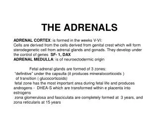
THE ADRENALS ADRENAL CORTEX: is formed in the weeks V-VI: Cells are derived from the cells derived from genital crest which will form sterodegenetic cell from adrenal glands and gonads. They develop under the control of genes SF- 1, DAX ADRENAL MEDULLA: is of neuroectodermic origin Fetal adrenal glands are formed of 3 zones: “definitive” under the capsulla (it produces mineralocorticoids ) of transition ( glucocorticoids) fetal zone has the most important area during fetal life and produces androgens - DHEA-S which are transformed within e placenta into estrogens zona glomerulosa and fasciculata are completely formed at 3 years, and zona reticularis at 15 years
Zona reticularis Zona fasciculata Zona glomerulosa
Pattern of steroidogenesis within the adrenal cortex • Glucocorticoid synthesis is produced into zona fasciculataCholesterol is converted into pregnenolone due to an enzyme that produces the clivage of lateral chain of cholesterol and StAR – steroidogenetic acute regulatory protein1.Pregnenolone is transformed into 17 hydroxi-pregnenolone due to enzyme 17 hydroxilase (C17)2. 17 hydroxi-pregnenolone in transformed into 17 hyroxiprogesterone due to 3β hydroxysteroid dehydrogenase3. 17 hyroxiprogesterone is transformed into 11 deoxy-cortisole by 21 hydroxilase4.11 deoxy-cortisole is converted into cortisole by 11 β hydroxilase
Mineralocorticoid biosynthesis 1. Cholesterol is converted into pregnenolone due to an enzyme that produces the clivage of lateral chain of cholesterol and StAR – steroidogenetic acute regulatory protein 2. Pregnenolone is transformed into progesterone due to enzyme 3β hydroxysteroid dehydrogenase 3. Progesterone is converted into deoxycorticosterone under the action of 21 hydroxilase 4. Deoxycorticosterone is convertes into corticosterone due to 11 β hydroxilase 5. Corticosterone is transformed into 18 hydroxi-corticosterone due to 18 hyroxilase (corticosterone methyl oxidase I) 6. 18 hydroxi-corticosterone is converted into aldosterone due to 18 hydrogenase (corticosterone methil oxidase II)
Adrenal failure Adrenal insufficiency is a disorder first described by Thomas Addison in 1855, which is characterized by impaired adrenocortical function and decreased production of glucocorticoids, mineralocorticoids and/or adrenal androgens. Adrenal insufficiency can be caused by diseases affecting the adrenal cortex (primary), the pituitary gland and the secretion of adrenocorticotropic hormone (ACTH) (secondary) or the hypothalamus and the secretion of corticotropic-releasing hormone (CRH) (tertiary).
Primary – due to adrenal failure Secondary – due to ACTH deficiency Tertiary – due to CRH deficiency Prevalence: • 39-60 new cases in 1 million /year • 60-70 % of cases are diagnosed between 30 – 50 de years • Sex ratio : F/M = 1,25 /1 in tuberculosis si 2,6 - 3/1 in autoimmune adrenal failure
Causes of Primary Adrenal Insufficiency • The etiology of primary adrenal insufficiency has changed over time. Prior to 1920, the most common cause of primary adrenal insufficiency was tuberculosis, while since 1950, the majority of cases have been ascribed to autoimmune adrenalitis, either in isolation or in the context of complex polyglandular syndromes
Causes of Primary Adrenal Insufficiency • Autoimmune adrenalitis: This condition is the result of an autoimmune process that destroys the adrenal cortex. Both humoral and cell-mediated immune mechanisms directed at the adrenal cortex are involved. The adrenals appear small and atrophic and the surrounding capsule is thickened. Antibodies that react with several steroidogenic enzymes, as well as all three zones of the adrenal cortex are detected in 60-75% of patients with autoimmune primary adrenal insufficiency, but only rarely in patients with other causes of adrenal insufficiency or normal subjects
Causes of Primary Adrenal Insufficiency . Considerable progress has been made in identifying genetic factors that predispose to the development of autoimmune adrenal insufficiency . In addition to the well-known association of the HLA genotype DR3/4-DQB1*0302 with type 1 diabetes mellitus and adrenal insufficiency, a strong susceptibility for the latter was also found for the DR3-DQ2/DRB1*0404-DQ8 genotype, which predicted early onset of primary adrenal insufficiency Approximately 50% of patients with autoimmune adrenal insufficiency have one or more other autoimmune endocrine disorders, whereas patients with the more common autoimmune endocrine disorders, such as type 1 diabetes mellitus, chronic autoimmune thyroiditis, or Graves' disease, rarely develop adrenal insufficiency. The combination of autoimmune adrenal insufficiency with other autoimmune endocrine disorders is referred to as the polyglandular autoimmune syndromes type I and II
Causes of Primary Adrenal Insufficiency • Primary Adrenal InsufficiencyAutoimmune (polyglandular failure) • Tuberculosis • Sarcoidosis, amyloidosis, hemochromatosis • Hemorrhage (meningococcemia, anticoagulants, trauma) • Fungal infections • Metastatic neoplasia/infiltration • Congenital adrenal hyperplasia • Congenital adrenal hypoplasia hypoplasia drenalis congenita • Congenital unresponsiveness to ACTH (glucocorticoid deficiency, ACTH resistance) • Adrenoleukodystrophy/Adrenomieloneuropathy • Aquired immynodeficiency syndrome • Bilateral adrenalectomy • Steroid synthesis inhibitors (e.g., metyrapone, ketoconazole, aminoglutethimide) • Adrenolytic agents (o,p’DDD, suramin) • Glucocorticoid antagonists (RU 486)
Causes of secondary and tertiary Adrenal Insufficiency • Secondary and Tertiary Adrenal Insufficiency • Following discontinuation of exogenous glucocorticoids or ACTH • Following the cure of Cushing’s syndrome • Pituitary and hypothalamic lesions • Tumors • Inflammation • Infections • Autoimmune lesions • Granulomatios infilitration • Trauma • Congenital aplasia, hypoplasia, dysplasia, ectopy • Pituitary-hypothalamic surgery • Pituitary-hypothalamic radiation • Pituitary-hypothalamic hemorrhage (apoplexy) • Acquired isolated ACTH deficiency • Familial corticosteroid-binding-globulin deficiency
Causes of Primary Adrenal Insufficiency • Infectious adrenalitis: Many infectious agents may attack the adrenal gland and result in adrenal insufficiency, including tuberculosis (tuberculous adrenalitis), disseminated fungal infections and HIV-associated infections, such as adrenalitis due to cytomegalovirus and mycobacterium avium complex . • Hemorrhagic infarction: Bilateral adrenal infarction caused by hemorrhage or adrenal vein thrombosis may also lead to adrenal insufficiency . The diagnosis is usually made in critically ill patients in whom a computed tomography (CT) scan of the abdomen shows bilateral adrenal enlargement. Several coagulopathies and the heparin-induced thrombocytopenia syndrome have been associated with adrenal vein thrombosis and hemorrhage, while the primary antiphospholipid syndrome has been recognized as a major cause of adrenal hemorrhage . Adrenal hemorrhage has been mostly associated with meningococcemia (Waterhouse-Friderichsen syndrome) and Pseudomonas aeruginosa infection).
Causes of Primary Adrenal Insufficiency • Adrenoleukodystrophy: This is an X-linked recessive disorder affecting 1 in 20.000 males . Adrenoleukodystrophy is characterized by spastic paralysis and adrenal insufficiency, usually beginning in infancy or childhood, and is caused by mutations in the ABCD1 gene, resulting in defective beta oxidation of very long chain fatty acids (VLCFAs) within peroxisomes. The abnormally high concentrations of VLCFAs in many organs, including the adrenal cortex, result in the clinical manifestations of this disorder.
Causes of Primary Adrenal Insufficiency • Drug-induced adrenal insufficiency: Drugs that may cause adrenal insufficiency by inhibiting cortisol biosynthesis, particularly in individuals with limited pituitary and/or adrenal reserve, include aminoglutethimide (antiepileptic), etomidate (anesthetic-sedative) , ketoconazole (antimycotic) and metyrapone. Drugs that accelerate the metabolism of cortisol and most synthetic glucocorticoids by inducing hepatic mixed-function oxygenase enzymes, such as phenytoin, barbiturates, and rifampicin can also cause adrenal insufficiency in patients with limited pituitary or adrenal reserve, as well as those who are on replacement therapy with glucocorticoids . Furthermore, some of novel tyrosine kinase-targeting drugs (e.g. sunitinib) have been shown in animal studies to cause adrenal dysfunction and • Adrenal hemorrhage in the context of a sepsis, anticoagulant treatment .
PATHOPHYSIOLOGY OF ADRENAL INSUFFICIENCY Pathophysiology of Primary Adrenal Insufficiency • In primary adrenal insufficiency, all the above mentioned causes result in gradual destruction of the adrenal cortex. However, the clinical manifestations of the condition appear when the loss of the adrenocortical tissue of both glands is higher than 90% . In the initial phase of chronic gradual destruction, the adrenal reserve is decreased and although the basal steroid secretion is normal, the secretion in response to stress is suboptimal. Consequently, any major or even minor stressor can precipitate an acute adrenal crisis. With further loss of adrenocortical tissue, even basal steroid secretion is decreased, leading to the clinical manifestations of the disease. Low plasma cortisol concentrations result in the increase of production and secretion of ACTH due to decreased negative feedback inhibition . The elevated plasma ACTH concentrations are responsible for the well-recognised hyperpigmentation observed in these patients. Pathophysiology of Secondary Adrenal Insufficiency • ACTH deficiency leads to decreased secretion of cortisol and adrenal androgens, while mineralocorticoid production remains normal. In the early stages, basal ACTH secretion is normal, while that of stress-induced is impaired . With further loss of basal ACTH secretion, there is atrophy of zonae fasciculata and reticularis of the adrenal cortex. Therefore, basal cortisol secretion is decreased but aldosterone secretion by the zona glomerulosa is preserved.
Adrenal insufficiency signs and symptoms • Chronic Primary Adrenal Insufficiency: Patients with chronic primary adrenal insufficiency may have symptoms and signs of glucocorticoid, mineralocorticoid, and androgen deficiency. By contrast, patients with secondary or tertiary adrenal insufficiency usually have normal mineralocorticoid function. The onset of chronic adrenal insufficiency is often insidious and the diagnosis may be difficult in the early stages of the disease.
Adrenal insuficiency signs and symptoms Clinical signs and symptoms occur when more than 90 % of adrenals is non functional. Glucocorticoid defficiency determines by feed-back an increased of pro-opio melanocortin and ACTH and MSH respectively . with skin pigmentation. Chronic adrenal insufficiency evolves in 4 stages: • aldosterone deficiency with increased plasma rennin activity. • subclinical cortisole deficiency with increased ACTH • low response of cortisole to SCTH stimulation test • Clinical signs and symptoms of the disease • Cortisole deficiency: hypoglycemia, lethargy, reducer apetite, anemia, depression. • Aldosterone deficiency: hyponatremia, reduced plasma volume , low blood pressure, reducered ability of kidney to respond to vasopressin and excrete free water.
Adrenal insufficiency signs and symptoms • The most common clinical manifestations of chronic primary adrenal insufficiency include: • General malaise, fatigue, weakness, • anorexia, weight loss, • nausea, vomiting, abdominal pain or diarrhea, which may alternate with constipation, • hypotension, • electrolyte abnormalities (hyponatremia, hyperkalemia, metabolic acidosis), • hyperpigmentation, autoimmune manifestations (vitiligo), • decreased axillary and pubic hair, and loss of libido and amenorrhea in women
Adrenal insufficiency signs and symptoms • Skin and mucosal pigmentation occurs on areas exposed to sun, exposed to pressure such as knuckles, toes, elbows and knees. Pigmentation of bucal mucosa and gums are preceded by generalized skin pigmentation. Increased pigmentation of palmar creases, nail beds, nipples, areolae, perivaginal and perianal mucosa is also found. Scars that have been formed after the onset of the diseas are also pigmented.