Download
1 / 19
190 likes | 245 Vues
Discover the layers of pericardium - fibrous and serous, their structures, and functions. Learn about pericardial fluid, its composition, role in heart health, and the importance of pericardial fluid analysis in diagnosing pericarditis and effusion.

E N D
Lecture on pericardium and pericardial fluid By Dr.Muhammadshahidsaeed
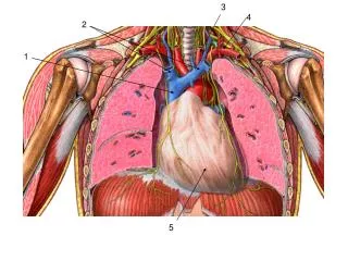
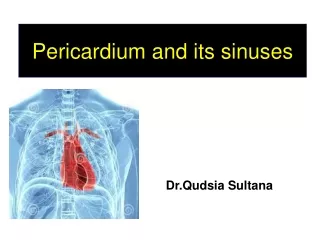
PERICADIUM • The membranous sac filled with serous fluid that encloses the heart and the roots of the aorta and other large blood vessels. • A superficial fibrous pericardium. • A deep two-layer serous pericardium: • The parietal layer lines the internal surface of the fibrous pericardium • The visceral layer or epicardium lines the surface of the heart • They are separated by the fluid-filled pericardial cavity.
Layers of pericardium • Fibrous Pericardium • • It is a sac made up of connective tissue fully surrounding the heart with out being attached to it • It is roughly conical in shape • It is superiorly connected with tunica adventitia of great vessels • Inferiorly it is connected with central tendon of diaphragm • Anteriorly it is separated from thoracic wall by lung & pleura, however some portion of it is in direct relation with left half of lower part of body of Sternum and left 4th &5th costal cartilages
Posteriorly it is related to esophagus descending thoracic Aorta & posterior part of mediastinal surface of both lungs • Serous Pericardium • •It is closed sac within fibrous pericardium having Visceral & Parietal layer • •The visceral layer of serous pericardium (epicardium) covers the surface of the heart • •It also reflects onto the great vessels
•From around the great vessels, the serous pericardium reflects to line the internal aspect of the fibrous pericardium as the parietal • layer of serous pericardium • Transverse Sinus • The transverse sinus is bounded anteriorly by the serous pericardium covering the posterior aspect of the pulmonary trunk and aorta, and posteriorly by the visceral pericardium covering the atria • The transverse pericardial sinus is especially important to cardiac surgeons.
• After the pericardial sac has been opened anteriorly, a finger can be passed through the transverse pericardial sinus posterior to the aorta and pulmonary trunk. • By passing a surgical clamp or placing a ligature around these vessels, inserting the tubes of a coronary bypass machine, and then tightening the ligature, surgeons can stop or divert the circulation of blood in these large arteries while performing cardiac surgery.
Oblique Sinus • The oblique sinus is bounded • a. anteriorly by the visceral layer of serous pericardium covering the left • atrium • b. posteriorly by the parietal layer of serous pericardium lining the fibrous pericardium, • c. superiorly and laterally by the reflection of serous pericardium around the four pulmonary veins and the superior and inferior venaecavae
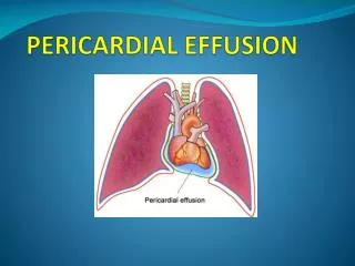
tamponade Cardiac • • Cardiac tamponade (heart compression) is due to • critically increased volume of fluid outside the heart but inside the pericardial cavity; e.g., due to stab wounds or from perforation of a weakened area of the heart muscle after heart attack (hemopericardium).
Pericardial fluid is a liquid that acts as a lubricant for the movement of the heart. It is found in small quantities between the two layers of thepericardium. Pericardial fluid is produced by mesothelial cells in the membranes and acts to reduce friction as the heart pumps
About 20-60 ml of pericardial fluid is present in the sac . The average amount of fluid is 15-30 ml. This is produce by the process of ultrafiltration with very low amount of proteins that is why it is transudate in nature. It is consisted of Water Sodium Chloride Magnesium Potassium LDH
The Proteins are present in the form of albumin globulin and fibrinogen but there concentration is less than that of plasma. • As for as the cells are concerned the following cells are present in the pericardial fluid. • Mesothelial cells • Lymphocytes • Granulocytes • Macrophages • Eosinophil • Basophil Lymphocyte are the cells which are present in abundance in the pericardial fluid.
Functions:- 1. lubricating the moving surface of heart .2. Stabilizing the heart.3. isolation of the heart from adjacent structures so that the inflammatory and neuplastic extension towards heart is inhabitant.4. limiting the heart dilatation during diastole.5. Prevention of cardiac hyperthrophy during cardiac overload.6. reducing right ventricular impulse work during left ventricular overload.7. Preservation of negative endothoraxic which is important for the filling of atria.8. The Nervous stimulation response and regulation of cardiac frequency and blood pressure.
Pericardial Fluid Analysis • Pericardial fluid analysis is used to help diagnose the cause of inflammation of the pericardium called pericarditis and/or fluid accumulation around the heart (pericardial effusion). There are two main reasons for fluid accumulation, and an initial set of tests (fluid protein or albumin level, cell count, and appearance) is used to differentiate between the two types of fluid that may be produced. • An imbalance between the pressure within blood vessels (which drives fluid out of the blood vessel) and the amount of protein in blood (which keeps fluid in the blood vessel) can result in accumulation of fluid (called a transudate. Transudates are most often caused by congestive heart failure or cirrhosis. If the fluid is determined to be a transudate, then usually no more tests on the fluid are necessary. • Injury or inflammation of the pericardium may cause abnormal collection of fluid (called an exudate). Exudates are associated with a variety of conditions and diseases and several tests, in addition to the initial ones performed, may be used to help diagnose the specific condition, including: • Infectious diseases – caused by viruses, bacteria, or fungi. Infections may originate in the pericardium or spread there from other places in the body. For example, pericarditis may follow a respiratory infection. • Bleeding – bleeding disorders and/or trauma can lead to blood in the pericardial fluid. • Inflammatory conditions – pericarditis may follow a heart attack, radiation treatment, or be part of autoimmune disorders such as rheumatoid arthritis and systemic lupus erythematosus. • Cancer – such as mesothelioma that has arisen in the pericardium or metastatic cancer that has spread to it.
Pericardial fluid analysis • Chest pain, sharp or sometimes dull, that may be relieved by bending forward • Coughing • Difficulty breathing • Fever • Fatigue • Changes in heart rhythm • Enlarged heart size on chest X-ray • Abnormal pericardial appearance on echocardiogram
Transudate VS Exudate fluid. • Transudate • Transudates are most often caused by either congestive heart failure or cirrhosis. Typical fluid analysis results include: • Physical characteristics—fluid appears clear • Protein or albumin level—low • Cell count—few cells are present • Exudate • Exudates can be caused by a variety of conditions and diseases. Initial test results may show: • Physical characteristics—fluid may appear cloudy • Protein or albumin level—high • Cell count—increased