Download
1 / 38
570 likes | 3.06k Vues
Pericardium and its sinuses. Dr.Qudsia Sultana. Heart location. Features of the heart. Function of Pericardium. Contents of pericardium. Heart with cardiac vessels, nerves Ascending aorta Pulmonary trunk Superior vena cava (lower half) Inferior vena cava (terminal part)

E N D
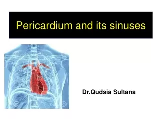
Pericardium and its sinuses Dr.Qudsia Sultana
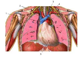
Contents of pericardium • Heart with cardiac vessels, nerves • Ascending aorta • Pulmonary trunk • Superior vena cava (lower half) • Inferior vena cava (terminal part) • Pulmonary veins (terminal parts)
Layers of Pericardium • Fibrous pericardium • Serous pericardium • Visceral layer • Parietal layer • Pericardial cavity between the 2 layers of serous pericardium – Capillary layer of fluid.
PERICARDIUM Heart projects into the serous pericardium from above and behind and reduces into a potential space. Hence serous pericardium consists of parietal and visceral layers.
Relations • Above – great vessels • Below – fuses with the central tendon of diaphragm. • Anterior –attached to sternum through sternopericardial ligaments. • Posterior –structures of psterior mediastinum • Laterally –mediastinal surfaces of left lung
Serous pericardium: • It is a closed serous sac lined by mesothelium present within the fibrous pericardium. • 2 layers – Parietal and Visceral • Pericardial cavity with capillary layer of fluid. • The continuity between parietal and visceral layers is established in the form of two tubes. • The reflections of serosal pericardium are in the form of two complex tubes…… The second tube around the SVC, IVC, and superior and inferior pulmonary veins One tube around the Asc. Aorta and Pulmonary trunk
Pericardial sinuses: • Transverse sinus • Oblique sinus
Transverse sinus: • Between the arterial and venous ends of primitive heart. • Formed by the degeneration of central cells of dorsal mesocardium • BOUNDARIES: Anteriorly :- Asc. Aorta & Pulmonary trunk. • Posteriorly :- SVC & upper border of the Lt atrium
Oblique sinus: • Cul-de-sac behind the left atrium • Formed by the absorption of pulmonary veins. • BOUNDARIES: • Right side :- Right pair of pulmonary veins & IVC • Left side :- left pair of pulmonary veins.
Pericardial sinuses Oblique sinus which lies behind the left atrium.
Fold of Marshall (fold of left vena cava): • Triangular fold of serous pericardium. • Extends from left pulmonary artery to left superior pulmonary vein. • Contains a fibrous ligament, a remnant of left duct of cuvier (left common cardinal vein).
BLOOD SUPPLY ARTERIAL SUPPLY VENOUS DRAINAGE
Nerve supply • Fibrous and Parietal layer— • Phrenic nerve– (sensory) • Pain sensitive • Visceral layer— • Vagus • Sympathetic– vasomotor • Non pain sensitive
Some interesting points….. • Surgical significance of transverse sinus:
Pericarditis, • Inflammation….. • Chest pain. Pericardial rub • Rough serous pericardium • Pericardial effusion • Inflammatory disease (pus..) • Non-inflammatory effusion • with congestive heart failure.
Cardiac Tamponade (pericardial tamponade):-- • Compression on the heart due to excessive fluid (blood, pus….) in the pericardial sac. • SYMPTOM: • Decreased cardiac output. • CAUSES: • Hypothyroidism, • Physical trauma (either penetrating trauma involving the pericardium or blunt chest trauma), • Pericarditis (inflammation of the pericardium), • Myocardial rupture.
Paracentesis of pericardial fluid: • Pericardial effusion • Drainage • 1) 5th or 6th ICS (lateral to apex beat) • 2) Angle (Xiphoid process & LT costal margin)