Protein Structure and Function
This comprehensive overview delves into protein structure and function, highlighting the significance of different structural levels, from primary (amino acid sequence) to quaternary (complex formations). Key processes such as protein characterization, modifications, and interactions including hydrophobic effects, ionic bonds, and hydrogen bonding are explored. The importance of structural motifs, experimental techniques for determining conformations, and tools for visualizing protein structures through resources like PDB and Cn3D are discussed, catering to students and researchers in bioinformatics and related fields.

Protein Structure and Function
E N D
Presentation Transcript
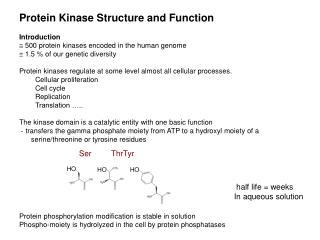
Protein Structure and Function 1, 2 , 3 , 4 Structure Viewing, interpreting structure Protein Characterization BIO520 Bioinformatics Jim Lund
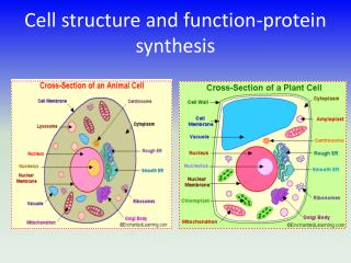
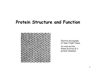
~100 to >10,000 aa Soluble Integral membrane proteins Membrane-associated Single domain, multiple domains singular, multimeric, large complex Protein variety DIVERSE
1 structure aa sequence 2 structure regular local folding 3 structure packing and overall folding 4 structure polypeptide:polypeptide complexes Protein Structure
Proteolysis/processing Residues Modified cysteine disulfides phosphorylation methylation Heteroatoms Metal ions, heme, cofactors…. Modifications
Dogma Sequence=Structure Similar Sequence Similar Function Kinetics vs Thermodynamics Chaperones
Restricted, but considerable rotation Different residuesdifferent , Regular , helix sheet , angles
Experimental , angles Hovmöller et al., 2002. Conformations of amino acids in proteins
An example Ramachandran plot Ideal a kinase
H-bonding orientation (dielectric) Hydrophobic effect nonpolar to core Ionic interaction + to -, (dielectric) Dipole effects helix, N to C (+ to -) Forces holding proteins together
right-handed aa preferences A,E,L,M (enriched) P,G,Y,S (less likely) 3.6 residues/turn helical wheel (amphipathic) dipole N to C -helix
Parallel or antiparallel N-to-C Mixed -sheets are rare: only ~20% of -sheets are mixed parallel/anti-parallel. “pleated” and “twisted” aa preferences -sheet
Length, conformation variable 2 aa hairpins common loops on surface diverge rapidly -turn, loops
hairpin (-loop- ) Helix-loop-helix Greek key (4 antiparallel , wrap) -- motif (-alpha helix-parallel ) Simple Super-2o Motifs Motifs-Domain Tertiary
Helix-loop-helixHelix-turn-helix DNA binding EF hand (Ca++ binding)
Cn3D (NCBI) .cn3 files (MMDB, NCBI structures) RasMol ProteinExplorer CHIME WWW compatible, animatable Jmol WWW compatible, animatable Protein Structure Viewers
MMDB, NCBI structures Cn3D format (ASN1) Protein Data Bank (PDB) PDB, Chime, other formats (http://www.pdb.org) Protein Structure databases