Download
1 / 15
150 likes | 343 Vues
Calcium Homeostasis and Signaling in Yeast Cells and Cardiac Myocytes. Jiangiun Cui, J.A. Kaandorp, P.M.A. Sloot Section Computational Science University of Amsterdam. Overview. General Introduction Calcium Networks in Yeast and Cardiac Myocytes Yeast Ca 2+ Dynamics & A Mathematical Model

E N D
Calcium Homeostasis and Signaling in Yeast Cells and Cardiac Myocytes Jiangiun Cui, J.A. Kaandorp, P.M.A. Sloot Section Computational Science University of Amsterdam
Overview • General Introduction • Calcium Networks in Yeast and Cardiac Myocytes • Yeast Ca2+ Dynamics & A Mathematical Model ---Control Block Diagram ---Mathematical Model ---Simulation Results • Ca2+-Calcineurin Network Controlling Heart Growth • Advantages of Calcium Signaling Research in Yeast • Conclusions and Future works • Acknowledgements & References
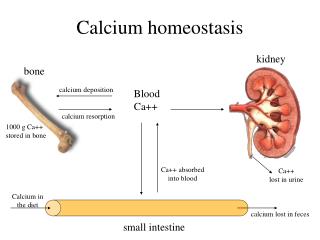
Ca2+, the Most Ubiquitous & Versatile Intracellular 2nd Messenger [1,7] • Ca2+ Homeostasis : -Dynamical balance -Basal level cytosolic Ca2+ : 50-200nM -Extracellular Ca2+ : ~1mM • Intracellular Ca2+ Signaling : -Various extracellular stimuli → Ca2+ transients, sparks, oscillations, puffs, etc. → downstream components → cell response (proliferation, muscle contraction, neurotransmitter release, programmed cell death, etc.)
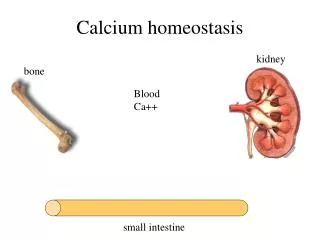
[2,3,5,8] Calcium Homeostasis Systems Cui and Kaandorp, Cell Calcium, 39: 337-348 (2006);Bers, Nature 415:198-205 (2002) Left: budding yeast cell Right: mammalian cardiac myocytes CaM: calmodulin; CaN: calcineurin; LTCC: L-Type Ca2+ channel; ATP: ATPase; NCX: Na+/Ca2+ exchanger; SR: sarcoplasmic reticulum; SERCA: SR Ca2+-ATPase; ATP: ATPase; RyR: ryanodine receptor; mRyR: mitochondrial ryanodine receptor; UP: mitochondrial uniporter; PLB: phospholamban; RaM: rapid-mode uptake pathway
Comparison of Two Systems [2,3,5,9] • Both systems have a set of calcium homeostasis and signaling toolkits composed of channels, pumps, exchangers and other relevant components (sensors like calmodulin, effectors such as calcineurin, etc.) • Calcium homeostasis process in cardiac myocytes is inseparable from the calcium signaling process related to excitation-contraction coupling.Sarcolemmal membrane potential plays critical role regulating sarcolemmal Leak, LTCC and sarcolemmal NCX, thuscalcium homeostasis/signaling in mammalian cardiac myocytes is tightly coupled with other ion homeostasis processes such as Na+ and K+ homeostasis • Phosphorylation of several key proteins (PLB, LTCC and RyR) by kinases such as protein kinase A (PKA) and calmodulin-dependent protein kinase II(CaMKII) play vital role in calcium homeostasis process in cardiac myocytes because of the short time duration of the heart beat (typically <1s)
Control Block Diagram (cch1 yvc1 mutant) [5] Cui et al., Cell Calcium, in press, (2008) (doi:10.1016/j.ceca.2008.07.005) [Caex]: extracellular Ca2+ concentration; Vol: the volume of the cytosol x(t): cytosolic Ca2+ concentration; Ca2+-bound calmodulin is assumed to inhibit the activity of both transporters M and X
Mathematical Model (composed of 3 equations) [5] Cui et al., Cell Calcium, in press, (2008) (doi:10.1016/j.ceca.2008.07.005) • The main equation: x’(t): the change rate of cytosolic free Ca2+ concentration JM,JX, JPmc1, JVcx1 and JPmr1 : the calcium ion flux through Transporter M, Transporter X, Pmc1, Vcx1 and Pmr1 respectively Vol’(t) :the change rate of cytosolic volume f: the calcium buffer effect constant Uptake kinetics of Pmc1,Vcx1 and Pmr1: Michaelis-Menten equation Uptake behavior of Transporters M and X: Michaelis-Menten kinetics with competitive inhibition by extracellular Mg2+ concentration
[5] Ca2+ Transient Curves under Hypertonic Shock Stimuli: extracellular Ca2+ shock (800mM) + Mg2+ challenge The necessity of having two Mg2+-sensitive influx pathways (Transporters M and X) rather than having only one Mg2+-sensitive influx pathway (i.e., Transporter X) was demonstrated by both theoretical analysis and optimal fitting to the experimental data (left graph) using hybrid optimization algorithm. The validity of the model was further confirmed by its ability of reproducing the experimentally determined effects of Vcx1 on cytosolic free Ca2+ dynamics and simulating the effects of extracellular Mg2+ removal
[4,10] Complex Calcium-Calcineurin Signaling Network Cui and Kaandorp, LNCS 5103: 110–119 (2008) CaM: calmodulin; MCIP: modulatory calcineurin-interacting protein NFAT: nuclear factor of activated T-cells; NFATP: phosphorylated NFAT Stress: hypertrophic stimuli; PO: pressure Overload; CaN*: activated CaN
[4] Transient Curves for CaN* Over-expression Modeling idea: complex network → 17 reactions + 1 process→ 28 equations
[4] Dual Roles of MCIP in Cardiac Hypertrophy Left: reported (HW/BW: heart weight/body weight; TG: transgenic) Right: simulated CaN* overexpression causes the dissociation of Complex2 by promoting MCIPPP→MCIPP→MCIP which associates with CaN* to inhibit its activity. Moreover, the feedback loop of MCIP expression controlled by NFAT contributes significantly to the inhibition. In the case of PO, activated BMP1 promotes MCIP→MCIPP→MCIPPP which associates 14-3-3 to relieve its inhibition on hypertrophic response.
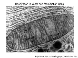
Why Study Yeast Calcium Signaling? [3,6,8] • Small cell size (1-7μm) , excluding the effect of diffusion • Unicellular, easy growth, relatively small genome size (6300 genes, about 1/5 of human genome) • A small set of relevant calcium transporters • Numerous mature technologies available (gene knockout, mass spectrometry, aequorin probing, etc.) • Most of the factors known in the yeast calcium homeostasis/signaling network are retained and operate similarly in mammalian cells including cardiac myocytes (Dolinski and Botstein 2007). For example, NFAT translocation in mammalian cardiac myocytes is strikingly similar as Crz1 translocation in yeast, MCIP signaling in cardiac myocytes is similar as Rcn signaling in yeast
[2,6,7,9,11] Conclusions • The extreme complexity of calcium homeostasis/signaling processes in cardiac myocytes arise from their built-in coupling with other ion homeostasis processes, the quite important spatial and stochastic effects, the great number of involved factors and the extremely sophisticated regulations (e.g., RyRs are regulated by numerous proteins) • Due to the universal conservation of yeast calcium homeostasis/signaling system across the eukaryotic kingdom, its understanding can be a shortcut to help understand the corresponding systems in mammalian cardiac myocytes and treat human diseases such as pathological cardiac hypertrophy and heart failure
Future Works • Mass-spectrometry-based proteomics can be a very powerful tool for searching the missing components, detecting and determining the protein interactions and quantifying the concentrations of proteins • Molecular genetic assays (e.g., yeast two-hybrid assay and gene knock-out technology) are important complementary methods to help elucidating the networksand provide powerful check for the validity of models • Effective collaborations among scientists who are proficient in genetics, proteomics and computational science via high-throughput experimental and computational methods through iterative systems biology procedure (model→experiment→model) are necessary
Acknowledgements & References -Thank the Dutch Science Foundation (NWO) and the European Commission(EC) for funding my research. -Thank Prof. Peter Sloot and Dr. Jaap Kaandorp for sustaining supports. -Thank Prof. Kyle Cunningham for valuable data & stimulating discussions. -Thank all the collaborators: Y. Fomekong Nanfack, Olufisayo O. Ositelu, Veronica Beaudry, Alicia Knight and Dr. Catherine M. Lloyd. -Thank Dr. Catherine M. Lloyd for translating two relevant models (Cui and Kaandorp 2006, 2008a) into CellML codes (see http://www.cellml.org/models/cui_kaandorp_2006_version03 • and http://www.cellml.org/models/cui_kaandorp_2008_version02, respectively) and include them into the CellML Model Repository. Main References: -1. Berridge et al., Nat. Rev. Mol. Cell Biol.4: 517-529 (2003). -2. Bers Nature415: 198-205 (2002). -3. Cui and Kaandorp,Cell Calcium39: 337-348 (2006). -4. Cui and Kaandorp, Lecture Notes in Computer Science 5103: 110–119 (2008). -5. Cui et al.,Cell Calcium (in press, 2008). (doi:10.1016/j.ceca.2008.07.005) -6. Dolinski and Botstein, Annu. Rev. Genet.41: 465-507 (2007). -7. Heineke and Molkentin, Nat. Rev. Mol. Cell Biol.7: 589-600 (2006). -8. Hilioti et al., Genes & Dev.18: 35 – 47 (2004). -9. Shannon et al., Biophys. J.87: 3351-3371 (2004). -10. Shin et al.,FEBS Letters580: 5965-5973 (2006). -11. Sobie et al., Biophys. J.83:59–78 (2002).