Circulation and Gas Exchange: Trading Places
Learn about the various exchange systems in organisms, including gills and vascular cavities. Understand the components and types of circulatory systems, such as open and closed systems. Discover the importance of double circulation and its benefits in maintaining high blood pressure.

Circulation and Gas Exchange: Trading Places
E N D
Presentation Transcript
Chapter 42 Circulation and Gas Exchange
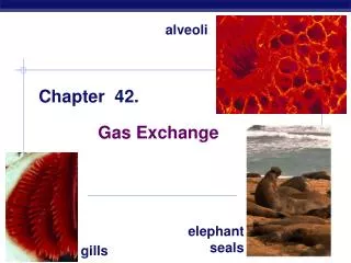
Overview: Trading Places • Every organism must exchange materials with its environment • Exchanges ultimately occur at the cellular level • In unicellular organisms, these exchanges occur directly with the environment • Gills are an example of a specialized exchange system in animals
Diffusion rates • The rate of diffusion from one place to another is proportional to square of the distance. • Because the rate of diffusion is so slow, there are two general solutions: • A body size and shape that keeps most cells in direct contact with the environment; • A circulatory system that moves fluid between each cell’s immediate surroundings and the tissues where exchange with the environment occurs.
1. What are vascular cavities? • In hydras and other cnidarians, a central gastrovascular cavity functions in digestion and in the distribution of substances throughout the body. • The body wall is only two cells thick and encloses the gastrovascular cavity • This cavity functions in both digestion and distribution of substances throughout the body • Some cnidarians, such as jellies, have elaborate gastrovascular cavities • Flatworms have a gastrovascular cavity and a large surface area to volume ratio
Fig. 42-2 Circular canal Mouth Pharynx Mouth Radial canal 5 cm 2 mm (a) The moon jelly Aurelia, a cnidarian (b) The planarian Dugesia, a flatworm
2. What three basic components does a circulatory system have? • More complex animals have either open or closed circulatory systems • Both systems have three basic components: • A circulatory fluid (blood or hemolymph) • A set of tubes (blood vessels) • A muscular pump (the heart)
3. Distinguish between open and closed circulatory systems. • In insects, other arthropods, and most molluscs, blood bathes the organs directly in an open circulatory system • In an open circulatory system, there is no distinction between blood and interstitial fluid, and this general body fluid is more correctly called hemolymph
3. Distinguish between open and closed circulatory systems. • In a closed circulatory system, blood is confined to vessels and is distinct from the interstitial fluid • Closed systems are more efficient at transporting circulatory fluids to tissues and cells • Higher blood pressures • Regulating distribution of blood to different organs
Fig. 42-3 Heart Heart Blood Hemolymph in sinuses surrounding organs Small branch vessels In each organ Interstitial fluid Pores Dorsal vessel (main heart) Tubular heart Auxiliary hearts Ventral vessels (a) An open circulatory system (b) A closed circulatory system
4. What is the total length of blood vessels in human adult? List the hierarchy of vessels in human circulatory system. • The total length of blood vessels in an average human adult is twice Earth’s circumference at the equator! • Humans and other vertebrates have a closed circulatory system, often called the cardiovascular system • The three main types of blood vessels are arteries, veins, and capillaries
Arteries branch into arterioles and carry blood to capillaries • Networks of capillaries called capillary beds are the sites of chemical exchange between the blood and interstitial fluid • Venules converge into veins and return blood from capillaries to the heart
Bony fishes, rays, and sharks have single circulation with a two-chambered heart • In single circulation, blood leaving the heart passes through two capillary beds before returning Gill capillaries Gill circulation Artery Ventricle Heart Atrium Systemic circulation Vein Systemic capillaries
5. Why is our circulatory system called a double circulation system? • Amphibian, reptiles, and mammals have double circulation • Oxygen-poor and oxygen-rich blood are pumped separately from the right and left sides of the heart in two distinct circuits, the pulmonary and system circuit
In reptiles and mammals, oxygen-poor blood flows through the pulmonary circuit to pick up oxygen through the lungs • In amphibians, oxygen-poor blood flows through a pulmocutaneous circuit to pick up oxygen through the lungs and skin • Oxygen-rich blood delivers oxygen through the systemic circuit • Double circulation maintains higher blood pressure in the organs than does single circulation
Fig. 42-5 Amphibians Reptiles (Except Birds) Mammals and Birds Lung and skin capillaries Lung capillaries Lung capillaries Right systemic aorta Pulmocutaneous circuit Pulmonary circuit Pulmonary circuit Atrium (A) Atrium (A) A A A A V V Ventricle (V) V V Left systemic aorta Left Right Left Right Right Left Systemic circuit Systemic circuit Systemic capillaries Systemic capillaries Systemic capillaries
Amphibians • Frogs and other amphibians have a three-chambered heart: two atria and one ventricle • The ventricle pumps blood into a forked artery that splits the ventricle’s output into the pulmocutaneous circuit and the systemic circuit • Underwater, blood flow to the lungs is nearly shut off
Reptiles (Except Birds) • Turtles, snakes, and lizards have a three-chambered heart: two atria and one ventricle • In alligators, caimans, and other crocodilians a septum divides the ventricle • Reptiles have double circulation, with a pulmonary circuit (lungs) and a systemic circuit
Mammals and Birds • Mammals and birds have a four-chambered heart with two atria and two ventricles • The left side of the heart pumps and receives only oxygen-rich blood, while the right side receives and pumps only oxygen-poor blood • Mammals and birds are endotherms and require more O2 than ectotherms
Fig. 42-6 Capillaries of head and forelimbs Superior vena cava 7 Pulmonary artery Pulmonary artery Capillaries of right lung Aorta 9 Capillaries of left lung 3 3 2 4 11 Pulmonary vein Pulmonary vein 5 1 Right atrium Left atrium 10 Right ventricle Left ventricle Inferior vena cava Aorta Capillaries of abdominal organs and hind limbs 8
6. Mammalian cardiovascular system, • The mammalian cardiovascular system meets the body’s continuous demand for O2 • Blood begins its flow with the right ventricle pumping blood to the lungs • In the lungs, the blood loads O2 and unloads CO2 • Oxygen-rich blood from the lungs enters the heart at the left atrium and is pumped through the aorta to the body tissues by the left ventricle • The aorta provides blood to the heart through the coronary arteries
Blood returns to the heart through the superior vena cava (blood from head, neck, and forelimbs) and inferior vena cava (blood from trunk and hind limbs) • The superior vena cava and inferior vena cava flow into the right atrium Animation: Path of Blood Flow in Mammals
Fig. 42-7 Pulmonary artery Aorta Pulmonary artery Right atrium Left atrium Semilunar valve Semilunar valve Atrioventricular valve Atrioventricular valve Right ventricle Left ventricle
7. Distinguish between systole and diastole. • The heart contracts and relaxes in a rhythmic cycle called the cardiac cycle • The contraction, or pumping, phase is called systole • The relaxation, or filling, phase is called diastole
Fig. 42-8-1 Semilunar valves closed AV valves open 0.4 sec 1 Atrial and ventricular diastole
Fig. 42-8-2 Atrial systole; ventricular diastole 2 Semilunar valves closed 0.1 sec AV valves open 0.4 sec 1 Atrial and ventricular diastole
Fig. 42-8 Atrial systole; ventricular diastole 2 Semilunar valves closed 0.1 sec Semilunar valves open AV valves open 0.4 sec 0.3 sec 1 Atrial and ventricular diastole AV valves closed 3 Ventricular systole; atrial diastole
The heart rate, also called the pulse, is the number of beats per minute • The stroke volume is the amount of blood pumped in a single contraction • The cardiac output is the volume of blood pumped into the systemic circulation per minute and depends on both the heart rate and stroke volume
Four valves prevent backflow of blood in the heart • The atrioventricular (AV) valves separate each atrium and ventricle • The semilunar valves control blood flow to the aorta and the pulmonary artery
8. What is “lub dup,” and what causes it? • The “lub-dup” sound of a heart beat is caused by the recoil of blood against the AV valves (lub) then against the semilunar (dup) valves • Backflow of blood through a defective valve causes a heart murmur
9. What is the sinoatrial node, and what is its function? • The sinoatrial (SA) node, or pacemaker, sets the rate and timing at which cardiac muscle cells contract • Impulses from the SA node travel to the atrioventricular (AV) node • At the AV node, the impulses are delayed and then travel to the Purkinje fibers that make the ventricles contract
Impulses that travel during the cardiac cycle can be recorded as an electrocardiogram (ECG or EKG) • The pacemaker is influenced by nerves, hormones, body temperature, and exercise
Fig. 42-9-5 3 1 2 Pacemaker generates wave of signals to contract. Signals are delayed at AV node. Signals pass to heart apex. Signals spread throughout ventricles. 4 SA node (pacemaker) AV node Purkinje fibers Bundle branches Heart apex ECG
Fig. 42-10 Artery Vein SEM Valve 100 µm Basal lamina Endothelium Endothelium Smooth muscle Smooth muscle Connective tissue Connective tissue Capillary Artery Vein Arteriole Venule 15 µm Red blood cell Capillary LM
10. Compare and contrast the structre of arteries, veins, and capillaries. • Capillaries have thin walls, the endothelium plus its basement membrane, to facilitate the exchange of materials • Arteries and veins have an endothelium, smooth muscle, and connective tissue • Arteries have thicker walls than veins to accommodate the high pressure of blood pumped from the heart • In the thinner-walled veins, blood flows back to the heart mainly as a result of muscle action
11. Does blood flow slower or faster in capillaries as compared to arteries? Why? • Physical laws governing movement of fluids through pipes affect blood flow and blood pressure • Velocity of blood flow is slowest in the capillary beds, as a result of the high resistance and large total cross-sectional area • Blood flow in capillaries is necessarily slow for exchange of materials
Fig. 42-11 5,000 4,000 Area (cm2) 3,000 2,000 1,000 0 50 40 Velocity (cm/sec) 30 20 10 0 120 Systolic pressure 100 80 Pressure (mm Hg) 60 Diastolic pressure 40 20 0 Aorta Veins Arteries Venules Arterioles Capillaries Venae cavae
12. Define vasoconstriction and vasodilation, and describe their effects on blood pressure. • Blood pressure is the hydrostatic pressure that blood exerts against the wall of a vessel • Blood pressure is determined by cardiac output and peripheral resistance due to constriction of arterioles • Vasoconstriction is the contraction of smooth muscle in arteriole walls; it increases blood pressure • Vasodilation is the relaxation of smooth muscles in the arterioles; it causes blood pressure to fall
Changes in Blood Pressure During the Cardiac Cycle • Systolic pressure is the pressure in the arteries during ventricular systole; it is the highest pressure in the arteries • Diastolic pressure is the pressure in the arteries during diastole; it is lower than systolic pressure • A pulse is the rhythmic bulging of artery walls with each heartbeat
Blood Pressure and Gravity • Blood pressure is generally measured for an artery in the arm at the same height as the heart • Blood pressure for a healthy 20 year old at rest is 120 mm Hg at systole and 70 mm Hg at diastole
Fig. 42-13-1 Pressure in cuff greater than 120 mm Hg Rubber cuff inflated with air 120 Artery closed
Fig. 42-13-2 Pressure in cuff greater than 120 mm Hg Pressure in cuff drops below 120 mm Hg Rubber cuff inflated with air 120 120 Artery closed Sounds audible in stethoscope
Fig. 42-13-3 Blood pressure reading: 120/70 Pressure in cuff greater than 120 mm Hg Pressure in cuff drops below 120 mm Hg Pressure in cuff below 70 mm Hg Rubber cuff inflated with air 120 120 70 Artery closed Sounds audible in stethoscope Sounds stop
Fainting is caused by inadequate blood flow to the head • Animals with longer necks require a higher systolic pressure to pump blood a greater distance against gravity • Blood is moved through veins by smooth muscle contraction, skeletal muscle contraction, and expansion of the vena cava with inhalation • One-way valves in veins prevent backflow of blood
Fig. 42-14 Direction of blood flow in vein (toward heart) Valve (open) Skeletal muscle Valve (closed)
Two mechanisms regulate distribution of blood in capillary beds: • Contraction of the smooth muscle layer in the wall of an arteriole constricts the vessel • Precapillary sphincters control flow of blood between arterioles and venules
Fig. 42-15 Thoroughfare channel Precapillary sphincters Capillaries Arteriole Venule (a) Sphincters relaxed Arteriole Venule (b) Sphincters contracted
The critical exchange of substances between the blood and interstitial fluid takes place across the thin endothelial walls of the capillaries • The difference between blood pressure and osmotic pressure drives fluids out of capillaries at the arteriole end and into capillaries at the venule end
Fig. 42-16 Body tissue INTERSTITIAL FLUID Capillary Net fluid movement out Net fluid movement in Direction of blood flow Blood pressure Inward flow Pressure Outward flow Osmotic pressure Arterial end of capillary Venous end
Fluid Return by the Lymphatic System • The lymphatic system returns fluid that leaks out in the capillary beds • This system aids in body defense • Fluid, called lymph, reenters the circulation directly at the venous end of the capillary bed and indirectly through the lymphatic system • The lymphatic system drains into veins in the neck • Lymph nodes are organs that filter lymph and play an important role in the body’s defense