Download
1 / 10
100 likes | 259 Vues
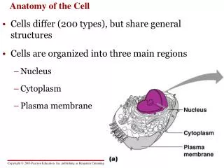
Anatomy of the Eukaryotic Cell. This lecture will be a brief review of eukaryotic structure and function. It is important that you have a clear understanding of these concepts so that you can move on to prokaryotic cellular structure and function. Structures external to cell wall.

E N D
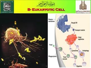
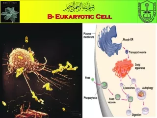
Anatomy of the Eukaryotic Cell This lecture will be a brief review of eukaryotic structure and function. It is important that you have a clear understanding of these concepts so that you can move on to prokaryotic cellular structure and function.
Structures external to cell wall • Some eukaryotic cells have appendages for motility. Motility is important for the organism to acquire nutrients and get away from toxic or harmful things in the environment. • Flagella on eukaryotic cells are few in number (1 or ) and long. • Flagellar movement is wavelike, like cracking a whip. • Cilia are also used for motility. • They are more numerous and much shorter than flagella (resemble hairs). They work like oars on a boat for move the cell from place to place. • Fewer types of cells have cilia as compared to those that have flagella.
Structures External to Cell Wall Continued • Glycocalyx: outermost covering found on most cells. • The glycocalyx is usually composed of sugars. • It functions to strengthen the cell surface. • It aids in adherence to other cells and surfaces. • It is important in signal reception between cells and the environment. • Generally the glycocalyx is categorized as either a capsule or a slime layer. • Ex. The glycocalyx is like the sugar coating on a jawbreaker. Underneath that first colored sugar coating there are many other layers.
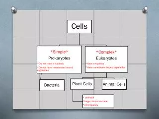
Cell Wall • Not all eukaryotes have a cell wall. • Plants and most algae have a cell wall composed of cellulose. Other cell walls are composed of pectin, mannans and minerals. • Fungi have cell walls composed of chitin. • Chitin is the same component that makes up the exoskeleton of crustaceans and insects. • The cell wall provides structure and shape to the cell. For example, plant cells look rectangular. It is the cell wall that holds the cell in that particular shape.
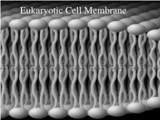
Cell/Plasma Membraneaka. Cytoplasmic Membrane • The cell membrane is a phospholipid bilayer. That means that each half of the membrane is composed of a phosphate group with 2 lipid chains attached to it. (See diagram on next page.) You can also find a diagram of a phophate group on page 44 of your textbook. • Within the lipid bilayer there are proteins. These proteins are often used to transport nutrients into the cell and waste outside of the cell. • The cell membrane also acts as a selectively permeable barrier for the transport of nutrients into and out of the cell. • In addition it encloses the cell’s organelles.
Structure of the Cell Memrane Outside Cell Phosphate Group Lipid or fat chain *Important! * The phosphorus loves the protons from the oxygen. It pulls the protons towards it and leaves the electrons on the outer edge of the oxygen. This creates a slight negative charge on the phosphate group. Ultimately the cell membrane is negatively charged. Transport protein within cell membrane R O - O P O Phosphate group looks like this Inside Cell O R
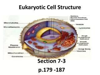
Structures inside cell membrane • The cell membrane encloses the cytoplasm. The cytoplasm is composed of a large amount of water. It is a substance in which various cellular components are found. • It contains the cell’s organelles. • It contains microfilaments, a network of protein strands used by phagocytes to form pseudopods. • It also contains microtubules, which are long, hollow tubes used for structure inside eukaryotic cells without a cell wall. • Microtubules also transport substances from one part of cell to another.
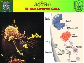
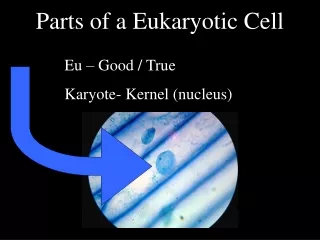
In addition, the cytoplasm houses the organelles. Organelles are structures within the cell that have specific shapes and specialized functions. • Organelles are found only eukaryotic cells. • The nucleus is the organelle that contains DNA, the genetic material of the cell. • The nucleus contains the nucleolus. It is the site for rRNA (ribosomal RNA) synthesis. • The Endoplasmic Reticulum (ER) is the factory for protein and lipid synthesis. • Rough ER has ribosomes attached to the outside. It’s job is to synthesize protein, (specifically proteins headed for the cell membrane). • Smooth ER has no ribosomes. It’s function is to synthesize lipids.
There are also free ribosomes, meaning that they are not bound to the ER, in the cytoplasm. Free ribosomes synthesize protein used inside cell. • The Golgi Complex (or Golgi Apparatus) is the post office for proteins. It packages and addresses the proteins for their final destination inside the cell. • The Mitochondria is the “power house” of the cell. It is responsible for ATP synthesis. • Aka. site of cellular respiration
Other types of organelles that some cells have are: • Lysosomes, whichstore digestive enzymes that are used by the cell to break down nutrients into units small enough to be utilized by the cell. • Vacuoles, which are temporary storage for proteins, sugars, organic acids, and inorganic ions. • Peroxisomes, which oxidize toxic substances into hydrogen peroxide. They also contain the enzyme catalase, that functions to safely decompose hydrogen peroxide into substances that are not harmful to the cell. • Now complete the homework for Lecture #2. It is due on Friday, September 8 at midnight.