Download

1 / 66
660 likes | 683 Vues
VITAL SIGNS. Module C. What are Vital Signs?. Temperature Pulse Respirations Blood Pressure Pain (considered the 5 th vital sign). When to measure vital signs?. On admission to health care facility In a hospital on regular hosp schedule or as MD ordered (q8hours, q4 hours, etc)

E N D
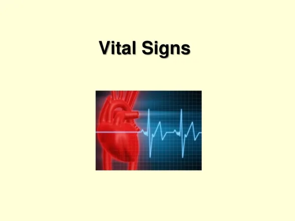
VITAL SIGNS Module C
What are Vital Signs? • Temperature • Pulse • Respirations • Blood Pressure • Pain (considered the 5th vital sign)
When to measure vital signs? • On admission to health care facility • In a hospital on regular hosp schedule or as MD ordered (q8hours, q4 hours, etc) • Before and after procedures (surgery, invasive diagnostic procedures) • Before, during, and after blood transfusions • When patient’s general condition changes (nursing judgment)
GUIDELINES FOR ASSESSMENT • Taken by nurse giving care • Equipment should be in good condition • Know baseline VS and normal range for pt and age group • Know pt’s medical history • Minimize environmental factors
GUIDELINES CONTINUED • Be organized in approach • Increase frequency of VS as condition worsens • Compare VS readings with the whole picture • Record accurately • Describe any abnormal VS
VS MUST BE ACCURATE • Both measuring and recording • VS vary according to pt’s illness/condition • Compare results with pt’s normal • Results are used to determine treatments, medications, diagnostic work, etc
REPORTING ABNORMAL VS • WHEN—grossly abnormal, return to normal, noted change for that pt • WHY—indicates change in metabolism or physiological function within the body • WHO—student reports to instructor, then TL, RN, Dr (follow chain of command) • HOW—orally to appropriate person, then document on chart
Body Temperature • Difference between heat produced by body processes and the heat lost to the external environment • Range 96.8 – 100.4 F (36 – 38 degree C) • Average for healthy young adults 98.6F or 37degrees C • No single temp is normal for all people
HEAT IS PRODUCED BY: • Metabolism • Increased muscle activity • Vasoconstriction • External sources
HEAT IS LOST BY: • Vasodilation • Convection • Radiation • Conduction • Evaporization
TEMP or FEVER? • TEMPERATURE—the measurement of heat in the body • FEVER—the measurement of heat in the body that is above normal for the individual
Adults- 96.8- 100.4 degree F Adult Avg 98.6 F Oral Adult Avg 99.5 F Rectal Adult Avg 97.7 F Ax Newborn range – 95.9- 99.5F Infants and children – same as adults Elderly – Avg 96.8F Normal Range Throughout Life Cycle
Frequently used terms: • Pyrexia or fever • Febrile • Hyperthermia • Hypothermia • Afebrile
FEVER—A DEFENSE MECHANISM • Indicator of disease in body • Pathogens release toxins • Toxins affect hypothalamus • Temperature is increased • Rest decreases metabolism and heat production by the body
PATTERNS OF FEVER • SUSTAINED- remains above normal with little change • RELAPSING – periods of febrile episodes interspersed with acceptable temp values • INTERMITTENT—varies from normal to above normal to below normal (may have a fairly predictable pattern) • REMITTENT—fever spikes and falls w/o a return to normal temp values
Age ( newborn- temp control mechanism immature, elderly- sensitive to temp changes) Exercise Hormonal level Circadian rhythm (temp normally changes 0.9 to 1.8 degree F /24hr Lowest 1-4AM Max-6PM ) Stress Environment Factors Affecting Body Temp
ORAL TEMPERATURE • Accessible • Dependable • Accurate • Convenient
RECTAL TEMPERATURE • Most reliable • MUST hold thermometer in place
AXILLARY TEMPERATURE • Safe • Non-invasive • Least accurate
TYMPANIC TEMPERATURE • Non-invasive • Safe • Accurate • Disadvantages • Excessive cerumen • Improper technique
AXILLARY TEMPERATUREIMPORTANT POINTS • AXILLA MUST HAVE ADEQUATE TISSUE & BE FREE OF PERSPIRATION • Not good method for persons with elevated temp • Used when cannot get oral or tympanic • Leave in place 10 minutes
ORAL TEMPERATURES • Wait 15-30 minutes after eating, drinking, chewing gum or smoking • If mouth breather-do not take orally • Leave in place 2 – 4 minutes with glass thermometer
TYMPANIC TEMPERATURES • Oral & tympanic readings will be same/ similar • Must direct probe toward TM (eardrum) • Follow instructions • Keep plugged in and on charger when not in use • Usually preferred method • Adults –pull pinna of ear up & back • Children under 3y/o-pull pinna of ear down & back
RECTAL TEMPERATURES • MOST accurate • MUST hold thermometer in place • Very high temp • Unconscious • Do not take rectal temp on clients with heart conditions • Leave in place 2-3 min with glass thermometer • Lubricate thermometer • DO Not take hand from thermometer while rectal in progress
NURSING DIAGNOSIS Hyperthermia> 100.4F Hypothermia <96.8F Risk for altered body temperature Ineffective Thermoregulation
Temperature Conversion • Temperature can be measured in Fahrenheit (F) or centigrade or Celsius (c) • To convert F to c, subtract 32 from F reading and multiply times 5/9. Ex. (104 F – 32) x 5/9 = 40 degree c • To convert c to F, multiply the c reading by 9/5 and add 32 to the product. Example (40 x 9/5) + 32 =104 F
Pulse • Pulse- is the palpable bounding of the blood noted at various points on the body. It is an indicator of circulatory status.
TERMS RELATED TO PULSE • Pulse—Rate, Rhythm, Quality • Pulse Deficit • Auscultate • Palpate • Tachycardia, Bradycardia
Temporal Carotid Apical Brachial Dorsalsis Pedis (Pedal) Radial and Apical are most common pulse sites used! Radial Ulnar Femoral Popliteal Posterior Tibial Pulse Sites
TECHNIQUE • Feel over BONY area • DO NOT use thumb • Use 2-3 fingers • DO NOT squeeze • Count 30 seconds if regular x 2 • Note Rate, Rhythm, Quality • If irregular, count for 1 full minute or take apical pulse for 1 minute.
APICAL-RADIAL PULSE • Requires 2 nurses • 1 nurse counts apical heart rate • 1 nurse counts radial pulse • BOTH count during the same 60 seconds • 1 nurse acts as timekeeper for both nurses
PULSE DEFICIT • Count apical-radial pulse • The difference is the PULSE DEFICIT • Apical pulse will always be the same or higher than the radial pulse if both are counted correctly • If the radial pulse is higher, one or both nurses counted incorrectly
Factors Affecting Pulse Rates • Exercise • Temperature • Emotions • Drugs • Hemorrhage • Postural Changes • Pulmonary Conditions
Variations of Pulse Rates • Tachycardia – Abnormally elevated pulse rate. (above 100 beats/ min) • Bradycardia – Abnormally slow pulse rate (less than 60 beats / min)
Pulse Rhythm • Regular – A regular interval of time occurs between each heartbeat or pulse felt. • Irregular – Interval interrupted by early, late, or missed beat.
Strength and Quality of Pulse • Pulse strength may be described as weak, strong, bounding, or thready. • PULSE GRADING (0-4 rating scale) • 0 – absent, not palpable • 1+ - diminished, barely palpable • 2+- easily palpable, normal pulse • 3+ - full, increased strength • 4+ - bounding, cannot be obliterated
Respirations • Mechanism the body uses to exchange gases between the atmosphere, blood, and the cells. Involves three processes: • Ventilation • Diffusion • Perfusion
PROCESS OF RESPIRATION • EXTERNAL RESPIRATION • Inhaled air enters lungs, at alveoli O2 crosses over to bloodstream • CO2 and other wastes cross over from bloodstream to alveoli and are exhaled • INTERNAL RESPIRATION • O2 carried in bloodstream crosses over to body cells • CO2 and other wastes from body cells cross over to the bloodstream
RESPIRATION • Chest Cavity—airtight vacuum with negative pressure • INSPIRATION—diaphragm contracts and pulls down, ribs move up, lungs fill with air • EXPIRATION—diaphragm relaxes and moves up, ribs move down, lungs expel air
COUNTING RESPIRATIONS • Count pulse first, then count respirations while holding wrist • Note rate, rhythm, quality, and character • Observe a full inspiration and expiration • Respiratory rates below 12 or greater than 20 require further assessment.
Counting Respirations cont. • If respirations regular, count respirations for 30 seconds and multiply times 2. • If irregular, less than 12 or greater than 20, count for 1 full minute. • Quality of respirations- assess movement of chest or abdominal wall- deep, normal, shallow • Deep- full expansion of lungs • Normal- normal • Shallow- limited expansion of lungs
Exercise Acute Pain Anxiety Smoking Body position Medications Neurological injury Age Environmental Temp Hemoglobin Function Factors Influencing Characteristics of Respirations
Blood Pressure • Force exerted on the walls of the artery. Created by the pulsing blood under pressure of the heart. • Systolic- Peak and maximum pressure of ejection of blood from the heart into the aorta. This is the top number. • Diastolic- The minimal pressure remaining the heart when the heart relaxes. This is the bottom number. • Recorded as a ratio Ex. 120/80 • Pulse pressure- Difference between the systolic and diastolic. ( 120/80 – Pulse pressure 40)