Download
1 / 25
250 likes | 273 Vues
Understand the anatomy of the eye and the role of rods and cones as visual receptors. Explore phototransduction processes from the eye to the brain, including GPCR cascade, signal transduction, and regulation mechanisms. Learn about the specialized properties of phototransduction and differences between rods and cones. Discover the retinal circuitry, connectivity in the retina, and the flow of visual information for a comprehensive understanding of how the eye sees.

E N D
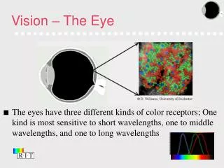
How the eye sees Last time Anatomy of the eye Rods and cones Visual receptors Color Vision This time Visual transduction Eye to brain 1
Rods and cones have different visual receptorsThe visual receptors are G protein-coupled receptors • seven transmembrane regions • hydrophobic/ hydrophilic domains • conserved motifs • chromophore stably attached to receptor (Schiff’s base Lys296 in TM7) • thermostable Nomenclature for visual receptors Receptor == GPCR, opsin Ligand == chromophore, retinal, pigment Receptor bound to ligand == rhodopsin 2
The visual cascade is a G protein-coupled cascade Rhodopsin Gtransducin phosophodiesterase cGMP to GMP close cGMP channels 4
Negative regulation of phototransduction Rhodopsin Gtransducin phosophodiesterase cGMP to GMP close cGMP channels Closing cGMP channels causes a decrease in Ca2+ Decrease in Ca2+ activates • Rhodopsin kinase === deactivate receptor • Guanylate cyclase === converts GTP to cGMP === opens cGMP channels Ca2+ independent deactivation also occurs 1. GTPase activating protein 7
Turning off rhodopsin Ca-dependent 8
Turning ON Guanylate Cyclase Ca-dependent, Decrease in Ca activates GC ENZYME REACTION ACTION on CHANNELS ACTIVATED BY Guanylate cyclase GTP--- cGMP opens cGMP channels GCAP (less Ca) Phosphodiesterase cGMP--- GMP closes cGMPchannels transducin 9
Mice without GAP cannot turn off light response quickly no GAP with GAP (wild-type) 11
Dark noise is very low 1 rhodopsin/minute 108 rhodopins/ photoreceptor 1000 years for all rhodopsins to turn over 14
High amplification increases signal size and reliability Rhodopsin Gtransducin phosophodiesterase cGMP to GMP close cGMP channels 1 100 100 100,000 ~1000 15
Properties of phototransduction • responds to 1 photon of light • responses are extremely reliable • high amplification of signaling • low dark noise • 1000s of discs maximize surface area of light detection • high concentration and thermostability of rhodopsin means high detection, low noise Photoreceptors are highly specialized to detect light! 16
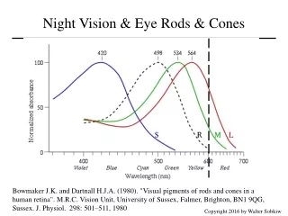
Phototransduction: Differences between rods and cones RodsCones Very sensitive to light 30x less sensitive to light each rhodopsin activates 30x less G proteins 17
The signaling pathway for Drosophila phototransduction Fastest GPCR cascade measured No amplification 19
Connectivity in the retina The Basic Retinal Circuit Back of eye 6. Pigment cells 1. Receptor Cells (rods and cones) 2. Bipolar Cells 3. Ganglion Cells 4. Horozontal Cells 5. Amacrine Cells Structure of the eye Front of eye 20
View of the retina Ramon y Cajal, Nobel 1906 21
Flow of visual information in the retina Vertical Connections Back of eye Front of eye back of eye Photoreceptor Cell---Bipolar Cell---Retinal Ganglion Cell---Brain Horozontal Connections Horozontal Cells- connect photoreceptors and bipolar cells Amacrine Cells- connect bipolar cells and retinal ganglion cells light 22
Retinal ganglion cells express melanopsin, are sensitive to light and project to the suprachiasmatic nucleus 25