Download
1 / 46
600 likes | 1.73k Vues
Fascia of the Abdomen. Chris van Zyl KHC. Outline. Peritoneum Embryology Anatomy Quick word on Fascia of the anterior abdominal wall Fascia around kidneys. Peritoneam. Serous membrane Divided into parietal and visceral

E N D
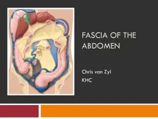
Fascia of the Abdomen Chris van Zyl KHC
Outline • Peritoneum • Embryology • Anatomy • Quick word on • Fascia of the anterior abdominal wall • Fascia around kidneys
Peritoneam • Serous membrane • Divided into parietal and visceral • Parietal peritoneum lines anterior, lateral and posterior walls peritoneal cavity • Visceral peritoneum lines all the organs that are intraperitoneal.
Embryology • Mesentries divide coelomic cavity into R + L halves • Upper abdomen • Stomach + gut suspended in the middle • Liver in the ventral mesentery • Spleen in dorsal mesentery
Embryology • Organs migrate in anticlockwise fashion • Liver lies on the right • Spleen on the left • Drags mesenteries into position they occupy in muturity
Peritoneal cavity Two main regions • Greater sac (general abdominal cavity) • Lesser sac (or omental bursa) • Communicate via foramen of Vinslow (epiploic foramen)
Course • Covers the inferior aspect of diaphragm and is reflected onto the liver and the abdominal part of the esophagus • After the liver is enclosed, it extends from the porta hepatis as a double layer (lesser omentum) to the lesser curvature of the stomach
Course • It encloses the stomach, reaches the greater curvature of stomach and extends as a double layer (greater omentum) down into the abdominal cavity,loops back up to the transverse colon. • From there the transverse mesocolon is formed, which joins the post abdominal wall on the anterior aspect of the pancreas
Course • Double layer devides into single layers • one which runs superiorly over the post abdominal wall and reflected onto the bare area of liver • The other runs inferiorly over the post abdominal wall to cover the pelvic organs and join the peritoneum of the anterior abdominal wall. • The post peritoneal layer of the post abdo wall is interrupted as it’s reflected from the duodenojejenal to ileocaecal junction to form the mesentery of the jejenum and ileum
Blood supply of the peritoneum To the parietal peritoneum • Lumbar vessels • Branches of the inferior and superior epigastric arteries • Musculophrenic arteries • Deep circumflex arteries To the visceral peritoneum • From the arteries supplying the appropriate viscera
Innervation of the peritoneum To the parietal peritoneum • From the nerves supplying the diaphragm and adjacent body wall - e.g. phrenic (C3-C5) and intercostal and subcostal nerves (T7-L1) To the visceral peritoneum • Sympathetic nerves innervating the appropriate viscera.
Peritoneam Anatomy devided into: • Peritoneal spaces • Peritoneal reflections including ligaments • Mesentry • Omenta
Peritoneal Spaces: • Peritoneal cavity devided by transverse colon and mesocolon into: • Supramesocolic compartment • Right and Left supramesocolic spaces • Inframesocolic compartment • Devided by root of smallbowel mesentery • Right and Left inframesocolic spaces
Right supramesocolic space Devided into: • Right subphrenic space • Diaphragmatic surface of R liver from falciform ligament medially to coronary ligament postero-infer • Right subhepatic space • Subdevided into ant + post spaces (Morrison’s pouch) • Communicates with R paracolic gutter + R infracolic space • Lesser sac
Left supramesocolic space Devided into: • Ant left perihepatic space • Post left perihepatic space (gastrohepatic recess) • Anterior left subphrenic spaces • Posterior left suphrenic spaces
Imaging RSP: R subphric S LSP: L subphrenic S LPC: L paracolic S LSs: Sup recess LS Lsi: Inf recess LS
Lesser Sac • Posterior Wall • Peritoneum over pancreas, left adrenal, upper pole of left kidney • Anterior Wall • Peritoneum over post. stomach, lesser omentum • Lateral Wall • Spleen, gastrosplenic, splenorenal ligaments • Communicates via epiploic foramen (of Winslow)
Lesser sac • Devided by gastropancreatic fold (left gastric artery) • Superior recess • Encloses caudate lobe • Inferior recess • Lies between stomach and pancreas • Left Gastric artery: Arrow • GHL: Gastro hepatic lig • SHS: Subhepatic S • LSs: Sup recess LS • Lsi: Inf recess LS
Foramen of Winslow • Post: IVC • Ant: free edge of lesser omentum containing portal vein, hepatic artery, CBD • Sup: Caudate lobe • Inf: 1st part of duodenum
Right inframesocolic compartment Bounded by: • Transverse mesocolon sup and to the R • Root of small bowel mesentery
Left inframesocolic space • Larger than right • In free communication with pelvis right to midline • Sigmoid colon and mesentery forms partial barier left of midline
Paracolic gutters • Peritoneal recesses lat to ascending + descending colon • Right continuous with right subhepatic/phrenic spaces • Left seperated by phrenicocolic ligament • Both communicates with pelvis
Pelvic peritoneal spaces/pouches • The rectouterine pouch/ Pouch of Douglas (in females) separating rectum from bladder • The rectovesical pouch (in males) separating the rectum from the bladder • The vesicouterine pouch (in females) separating bladder from uterus
Peritoneal reflections • 8 Legaments • 4 Mesentries • 2 Omenta
Peritoneal ligaments • Right coronary ligament • Reflection of peritoneum from diaphragm to post surface of R liver lobe • Bare area between two layers of this ligament • Left coronary ligament • Continuation of this peritoneal reflection to the left
Peritoneal ligaments • Gastrosplenic ligament • Continuous with greater omentum • From greater curve of stomach to spleen. • Contains gastroepiploic vessels + short gastric vessels • Falciform ligament • From anterosup surface of liver to diaphragm + ant abdominal wall • Carries lig Teres (obliterated umbilical vein) in free edge • Continues with fissure for lig venosum
Peritoneal ligaments • Phrenicocolic ligament • From splenic flexure to diaphragm • Continuous with transverse mesocolon + splenorenal lig • Splenorenal ligament • From pancreatic tail to splenic hilus • Transmits splenic vessels
Peritoneal ligaments • Hepatoduodenal ligaments • From flexure between 1st + 2nd part of duodenum to porta hepatis • Transports portal triad • Duodenocolic ligament • From R colic flexure to descending duodenum • Continuous with transverse mesocolon
Broad ligament of the uterus • Wide fold of peritoneum that connects the sides of the uterus to the walls and floor of the pelvis
Broad ligament of the uterus • Subcomponents: • Mesometrium- the mesentery of the uterus; the largest portion of the broad ligament • Mesosalpinx- the part that surrounds the Fallopian tube • Mesovarium- the part that connects the anterior surface of the ovary to the remainder of the broad ligament.
Mesentries • Small bowel mesentry • Broad fan shaped fold • Conects jejunum + ileum to post abdominal wall • Extends from duodenojejunal flexure (Left of L2) to upper part of R SI joint • Root continuous with L + R ant pararenal spaces • Contains jejunal, ileal branches of SMA and accompanying veins, nerves, lymphatics
Mesentries • Transverse mesocolon • Connects transvers colon to post abdominal wall • Pass from ant head and body of pancreas to post colon • Upper layer adherent to greater omentum • Contains middle colic vessels, nerves + lymphatics
Mesentries • Sigmoid mesocolon • Attaches sigmoid colon to pelvic wall • Line of attachment inverted V with apex near devision of L common iliac artery • Leftlimb descends to L psoas, R into pelvis • Contains sup rectal vessels • Mesoappendix • Attached to lower end of small bowel mesentry
Omenta • Greater and Lesser
Greater Omentum Hangs from the greater curve of the stomach and loops down in front of the intestines before curving back upwards to attach to the transverse colon, thus 4 layers of peritoneum Continuous with: • Gastrocolic ligament: largest component • Gastrosplenic ligament: to splenic hilus • Gastrophrenic ligament
Lesser Omentum Attached to the lesser curvature of the stomach, prox duodenum and the liver Subdivided into: • Gastrohepatic ligament • connects the left lobe of the liver to the lesser curvature of the stomach • Hepatoduodenal ligament • free edge of the omentum, extends to porta hepatis and ligamentum venosum • Contains gastric artery, coronary vein, left gastric nodal chain
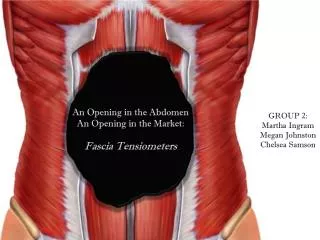
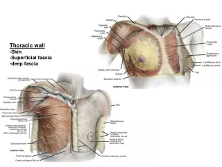
Fascia of anterior abdominal wall • Superficial fascia • Superficial layer • Deep layer • Transversalis fascia • 2: Superficial layer • 3: Deep layer • 8: Transversalis fascia
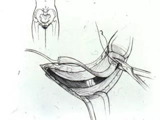
Superficial fascia • Single layer superiorly containing variable ammounts of fat • Inferiorly devides into superficial and deep layers • Superficial layer (Fascia of Camper): • Passes over inguinal ligament • Continuous with superficial fascia of the thigh • Continues in the scrotum as the fascia of Dartos
Superficial fascia • Deepy layer (Fascia of Scarpa): • Connected to aponeurosis of external oblique laterally + linea alba/ symphisis pubis medially • Passes over inguinal ligamnet inferolaterally to fuse with fascia of the thigh • Forms superfiscial perineal fascia inferomedially (Fascia of Colles)
Transversalis fascia • Lies between transverse abdominis and extraperitoneal fat • Continuous with inferior diaphragmatic fascia • Fuses with thoracolumbar fascia posteriorly • Spermatic cord/Round ligament passes through transversalis fascia at deep inguinal ring • becomes internal spermatic fascia
Fascia around kidneys • Renal fascia situated around the perirenal fat • Devided into: • Anterior fascia (Gerota’s) • Continuous with R inf coronary ligament • Posterior fascia (Zuckerkandl’s) • Continous with diaphragmatic fascia and iliacus fascia • Fused laterally as conal fascia • Continuous with transversalis fascia • Fused medially with sheeths of aorta + IVC
References: • Applied Radiological Anatomy • Paul Butler, Adam W. M. Mitchell, Harold Ellis • Anatomy for Diagnostic Imaging • Stephanie Ryan, Michelle McNicholas, Stephen Eustace • Third Edition