Download
1 / 94
950 likes | 1.55k Vues
Chapter 23. The Gastrointestinal Tract. Learning Objectives (1 of 2). Identify major types of cleft lip and cleft palate deformity Explain pathogenesis and prevention of dental caries and periodontal disease

E N D
Chapter 23 The Gastrointestinal Tract
Learning Objectives (1 of 2) • Identify major types of cleft lip and cleft palate deformity • Explain pathogenesis and prevention of dental caries and periodontal disease • Describe common congenital anomalies of the GIT, clinical manifestations, diagnosis, treatment • Describe three most common lesions of the esophagus that lead to esophageal obstruction • Explain pathogenesis, complications, and treatment of peptic ulcer • Describe types and clinical manifestations of acute and chronic enteritis
Learning Objectives (2 of 2) • Differentiate acute appendicitis and Meckel’s diverticulitis in terms of pathogenesis, clinical manifestations, and treatment • Describe pathogenesis of diverticulitis and the role of diet in its development • Discuss causes, clinical manifestations, complications • Intestinal obstruction • Colon cancer • Diverticulosis
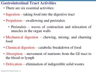
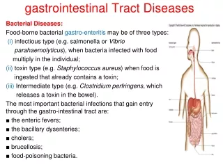
Gastrointestinal Tract • Digestion and absorption of food • Oral cavity • Esophagus, stomach, small and large intestines, anus
Cleft Lip and Cleft Palate • Embryologically, face and palate formed by coalescence of cell masses that merge to form facial structures • Palate formed by two masses of tissues that grow medially and fuse at midline to separate as nose and mouth • Maldevelopment leads to defects • 1 per 1000 births • Multifactorial inheritance pattern • Surgical correction (cheiloplasty) • Cleft lip: soon after birth • Cleft palate: 1 to 2 years of age followed by speech therapy to correct nasal speech
Types of cleft lip and palate abnormalities viewed from below
A complete bilateral cleft lip and cleft palate with anterior protrusion of tissues between clefts
The same child at 18 months after surgical correction of the defect
Abnormalities of Tooth Development • Teeth: specialized structures that develop in tissues of the jaws • Two sets • Temporary or deciduous teeth (20 teeth) • Permanent teeth (32 teeth) • Missing teeth or extra teeth: common abnormality • Enamel forms at specific times during embryologic period • Tetracycline: administered during enamel formation causes permanent yellow-gray to brown discoloration of the crown
Incisors Incisors Central (7 yr) Central (6–8 mo) Lateral (8 yr) Lateral (8–10 mo) Canine (eyetooth) (11 yr) Canine (eyetooth) (16–20 mo) Premolars (bicuspids) Molars First molar (10–15 mo) First premolar (11 yr) Deciduous (milk) teeth Second molar (about 2 yr) Second premolar (12–13 yr) Molars First molar (6–7 yr) Second molar (12–13 yr) Third molar (wisdom tooth) (17–25 yr) Permanent teeth (a) Figure 23.10a
Enamel Dentin Crown Dentinal tubules Pulp cavity (contains blood vessels and nerves) Neck Gingiva (gum) Cementum Root canal Root Periodontal ligament Apical foramen Bone Figure 23.11
Dental Caries and Periodontal Disease • Oral cavity: diverse collection of aerobic and anaerobic bacteria that mix with saliva, forming sticky film on teeth (dental plaque) • Plaque + action of bacteria result in tooth decay (caries) • Dental cavity: loss of tooth structure from bacterial action • Gingivitis: inflammation of the gums due to masses of bacteria and debris accumulating around base of teeth • Periodontal disease: inflammation extends to tissues that support teeth; forms small pockets of infection between teeth and gums • Two types: gingivitis and periodontitis
Tooth and Gum Disease • Dental caries (cavities): gradual demineralization of enamel and dentin • Dental plaque (sugar, bacteria, and debris) adheres to teeth • Acid from bacteria dissolves calcium salts • Proteolytic enzymes digest organic matter • Prevention: daily flossing and brushing
Tooth and Gum Disease • Gingivitis • Plaque calcifies to form calculus (tartar) • Calculus disrupts the seal between the gingivae and the teeth • Anaerobic bacteria infect gums • Infection reversible if calculus removed
Tooth and Gum Disease • Periodontitis • Immune cells attack intruders and body tissues • Destroy periodontal ligament • Activate osteoclasts • Consequences • Possible tooth loss, promotion of atherosclerosis and clot formation in coronary and cerebral arteries
Stomatitis • Inflammation of the oral cavity • Causes • Irritants: alcohol, tobacco, hot or spicy foods • Infectious agents: Herpes virus, Candida albicans fungus, bacteria that cause trench mouth
Carcinoma of the Oral Cavity • Arises from squamous epithelium • Lips • Cheek • Tongue • Palate • Back of throat
Esophagus (1 of 3) • Muscular tube that extends from pharynx to stomach with sphincters at both upper and lower ends • Upper sphincter relaxes to allow passage of swallowed food • Lower (gastroesophageal or cardiac) sphincter relaxes to allow passage of food to the stomach • Diseases • Failure of cardiac sphincter to function properly • Tears in lining of esophagus from retching and vomiting • At gastroesophageal junction from repetitive, intermittent, vigorous contractions that increase intraabdominal pressure • Esophageal obstruction from carcinoma, food impaction, or stricture
Bolus of food Tongue Uvula Pharynx Bolus Epiglottis Epiglottis Glottis Trachea Bolus Esophagus 1 2 3 Upper esophageal sphincter iscontracted. During the buccal phase, thetongue presses against the hard palate,forcing the food bolus into the oropharynxwhere the involuntary phase begins. The uvula and larynx rise to prevent foodfrom entering respiratory passageways. Thetongue blocks off the mouth. The upperesophageal sphincter relaxes, allowing foodto enter the esophagus. The constrictor muscles of thepharynx contract, forcing foodinto the esophagus inferiorly. Theupper esophageal sphinctercontracts (closes) after entry. Relaxed muscles 4 5 Food is movedthrough the esophagusto the stomach byperistalsis. The gastroesophagealsphincter opens, and foodenters the stomach. Relaxedmuscles Circular musclescontract Bolus of food Longitudinal musclescontract Gastroesophagealsphincter closed Gastroesophagealsphincter opens Stomach Figure 23.13
Relaxed muscles 4 Food is moved throughthe esophagus to thestomach by peristalsis. Circular musclescontract Bolus of food Longitudinal musclescontract Gastroesophagealsphincter closed Stomach Figure 23.13, step 4
Esophagus (2 of 3) • Symptoms • Difficulty swallowing (dysphagia) • Substernal discomfort or pain • Inability to swallow (complete obstruction) • Regurgitation of food into trachea • Choking and coughing • Two major disturbances of cardiac sphincter • 1. Cardiospasm: sphincter fails to open properly due to malfunction of nerve plexus; esophagus becomes dilated proximal to constricted sphincter from food retention • Treatment: periodic stretching of sphincter; surgery • 2. Incompetent cardiac sphincter: sphincter remains open; gastric juices leak back into esophagus
Esophagus (3 of 3) • Complications of incompetent cardiac sphincter • Reflux esophagitis: inflammation • Ulceration and scarring of squamous mucosal lining • Barrett’s esophagus: glandular metaplasia; change from squamous to columnar epithelium; ↑risk for cancer -a disorder in which the lining of the esophagus is damaged by stomach acid. • Esophageal obstruction • Carcinoma: can arise anywhere in esophagus • Tumor narrows lumen of esophagus, infiltrates surrounding tissue, invades trachea (tracheoesophageal fistula) • Food impaction: distal part • Stricture: from scar tissue due to necrosis and inflammation from corrosive chemicals such as lye
Gastric mucosal tear caused by retching and vomiting Mallory–Weiss syndrome or gastro-esophageal laceration syndrome refers to bleeding from tears (a Mallory-Weiss tear) in the mucosa at the junction of the stomach and esophagus, usually caused by severe retching, coughing, or vomiting.
Liver Gallbladder Lesser omentum Stomach Duodenum Transverse colon Small intestine Cecum Urinary bladder (b) Figure 23.30b
Surface epithelium Mucosa Lamina propria Muscularis mucosae Submucosa (contains submucosal plexus) Oblique layer Muscularis externa (contains myenteric plexus) Circular layer Longitudinal layer Serosa Stomach wall (a) Layers of the stomach wall (l.s.) Figure 23.15a
Gastric pits Surface epithelium (mucous cells) Gastric pit Mucous neck cells Parietal cell Chief cell Gastric gland Enteroendocrine cell (b) Enlarged view of gastric pits and gastric glands Figure 23.15b
Pepsinogen Pepsin HCl Mitochondria Parietal cell Chief cell Enteroendocrine cell (c) Location of the HCl-producing parietal cells and pepsin-secreting chief cells in a gastric gland Figure 23.15c
Blood capillary Chief cell Stomach lumen CO2 CO2 + H2O H+-K+ ATPase Carbonic anhydrase H2CO3 H+ H+ K+ K+ HCO3– HCI Parietal cell Alkaline tide HCO3– Cl– Cl– Cl– l HCO3–- Cl– antiporter Inter- stitial fluid Figure 23.18
Acute Gastritis • Inflammation of the gastric lining • Self-limited inflammation of short duration • May be associated with mucosal ulceration or bleeding • From nonsteroidal anti-inflammatory drugs (NSAID) that inhibit cyclooxygenase (COX) enzyme: aspirin, ibuprofen, naproxen • COX-1: promotes synthesis of prostaglandin that protects gastric mucosa • COX-2: promotes synthesis of prostaglandin that mediate inflammation • Drugs that selectively inhibit COX-2 increase risk for heart attack and stroke • Alcohol: a gastric irritant; stimulates gastric acid secretion
H. Pylori Gastritis (1 of 2) • Small, curved, gram-negative organisms that colonize surface of gastric mucosa • Grow within layer of mucus covering epithelial cells • Produce urease that decomposes urea, a product of protein metabolism, into ammonia • Ammonia neutralizes gastric acid allowing organisms to flourish; organisms also produce enzymes that break down mucus layer
H. Pylori Gastritis (2 of 2) • Common infection that increases with age (50% by age 50) • Spreads via person-to-person through close contact and fecal-oral route • Increased risk of gastric carcinoma: intestinal metaplasia • Increased risk of malignant lymphoma (mucosa-associated lymphoid tissue, MALT)
Bacteria Mucosa layer of stomach (b) H. pylori bacteria (a) A gastric ulcer lesion Figure 23.16
Peptic Ulcer • Pathogenesis • Digestion of mucosa due to increased acid secretions and digestive enzymes (gastric acid and pepsin) • Helicobacter pylori injures mucosa directly or through increased acid secretion by gastric mucosa • Common sites: distal stomach or proximal duodenum • Complications: hemorrhage, perforation, peritonitis, obstruction from scarring • Treatment • Antacids: block acid secretion by gastric epithelial cells • Antibiotic therapy: against H. pylori • Surgery if medical therapy fails
Bacteria Mucosa layer of stomach (b) H. pylori bacteria (a) A gastric ulcer lesion Figure 23.16
Gastric ulcer, eroded a blood vessel at base of ulcer causing profuse bleeding Large, chronic duodenal ulcer
Carcinoma of the Stomach • Manifestations • Vague upper abdominal discomfort • Iron-deficiency anemia (chronic blood loss from ulcerated surface of tumor) • Diagnosis: biopsy by means of gastroscopy • Treatment: surgical resection of affected part, surrounding tissue and lymph nodes • Long-term survival: relatively poor; often far-advanced at time of diagnosis
Inflammatory Diseases of the Intestines • Acute enteritis • Intestinal infections; common; of short duration • Nausea, vomiting, abdominal discomfort, loose stools • Chronic enteritis: less common, more difficult to treat • Regional enteritis or Crohn’s disease: distal ileum • Chronic inflammation and ulceration of mucosa with thickening and scarring of bowel wall • Inflammation may be scattered with normal intervening areas or “skip areas” • Treatment: drugs and possible surgical resection of affected part of bowel
Ulcerative Colitis (1 of 2) • Ulcerative colitis: large intestines and rectum • Inflammation is limited to mucosa, bowel not thickened unlike in Crohn’s • Frequently begins in rectal mucosa and spreads until entire colon is involved • Complications • Bleeding; bloody diarrhea • Perforation: from extensive inflammation with leakage of intestinal contents into peritoneal cavity • Long-standing disease may develop cancer of colon and/or rectum
Ulcerative Colitis (2 of 2) • Treatment • Symptomatic and supportive measures • Antibiotics, corticosteroids to control flare-ups • Immunosuppressive drugs • Surgical resection
Inflammatory Diseases of the Intestines (1 of 3) • Antibiotic-associated colitis: broad-spectrum antibiotics destroy normal intestinal flora • Allows growth of anaerobic spore-forming bacteria, Clostridium difficile not inhibited by antibiotic taken • Organisms produce toxins causing inflammation and necrosis of colonic mucosa • Diarrhea, abdominal pain, fever • Diagnosis: stool culture, toxin in stool • Treatment: stop antibiotic treatment; give vancomycin or metronidazole • Drugs that decrease intestinal motility will prolong illness
Inflammatory Diseases of the Intestines (2 of 3) • Appendicitis: most common inflammatory lesion of the bowel • Narrow caliber of appendix may be plugged with fecal material • Secretions of appendix drain poorly, create pressure in appendiceal lumen, compressing blood supply • Bacteria invade appendiceal wall causing inflammation • Manifestations • Generalized abdominal pain localizing in right lower quadrant; rebound tenderness; rigidity • Treatment: surgery
Inflammatory Diseases of the Intestines (3 of 3) • Meckel’s diverticulum • Outpouching at distal ileum, 12-18 inches proximal to cecum • From persistence of a remnant of the vitelline duct, narrow tubular channel connecting small intestine with yolk sac embryologically • Found in 2% of population; usually asymptomatic • May become infected causing features and complications similar to acute appendicitis • Lining may consist of ectopic acid-secreting gastric mucosa and may cause peptic ulcer
Regional enteritis, mucosa ulcerated and covered with inflammatory exudate