Respiratory Symptoms & Signs
1.05k likes | 1.64k Vues
Respiratory Symptoms & Signs. Chief complaints(CC): Presenting complaints and duration. Present Illness(PI):. A:The immediate history that brought the patient to the hospital B:Background history of disease leading to the immediate history

Respiratory Symptoms & Signs
E N D
Presentation Transcript
Chief complaints(CC): Presenting complaints and duration.
Present Illness(PI): A:The immediate history that brought the patient to the hospital B:Background history of disease leading to the immediate history C:Significant positive and negative data that might give clues useful in differential diagnosis
Habits: A:Use alcohol,tobacco…… B:Sexual habits Allergies: Hay fever,asthma,drugs
Common symptoms • Cough • Expectoration • Hemoptysis • Chest pain • Cyanosis • Dyspnea
What is cough? • A complex reflex arc. • A defense mechanism. • A factor in the spread of infection. • A common symptom. • A means of providing cardiopulmonary resuscitation.
Cough (a protective reflex): causes • Respiratory diseases___ the most common causes • Airway agents • Bronchitis, bronchiectasis, asthma, endobronchial tuberculosis, tumor, pharyngitis • Lung agents • Infection, edema, fibrosis, tumor • Cardiovascular diseases • pulmonary edema, pulmonary embolism
Cough : manifestations • Characteristics • Dry cough (non-sputum: non-infectious) • Productive cough (sputum: infectious, edema) • Attack • Time • season • Tone • Hoarseness • Brassy
Cough: accompany symptoms • Fever (infection) • Chest pain (infection, tumor, pleurisy, pneumothorax, pulmo embolism) • Dyspnea • Hemoptysis (bronchietasis, tuberculosis, tumor) • Bulk pus sputum (bronchietasis, lung abscess) • Wheezing (asthma, foreign body) • Clubbing fingers (bronchietasis, lung cancer, chronic lung abscess)
The Duration of Cough • Cough can be divided into 3 categories • - Acute (<3 weeks) • - Subacute (3-8 weeks) • - Chronic (>8 weeks) • Estimating the duration is the 1st step in narrowing the list of potential causes.
Most Common Causes of Acute Cough • URT Infections • - Common Colds • “Acute bronchitis” • Acute Bacterial Sinusitis • Bordetella pertussis Infection in Selected Communities • Exacerbations of Chronic Bronchitis • Allergic Rhinitis • Environmental Irritant Rhinitis
Commonest Causes of Subacute Cough After a Respiratory Infection • Postinfectious Cough • - B. pertussis infection • Bacterial sinusitis • Exacerbation of a pre-existing disease • - Asthma • - Chronic Bronchitis • Helpful Hints: • Pertussis is likely with cough-vomit syndrome with or without whoop • Consider all 3 when cough has a biphasic course
Summary of Results of the Diagnostic Evaluation of Chronic Cough • Chronic cough is often simultaneously due to more than 1 condition (18-93% of the time). • - It has been due to 3 diseases up to 42% of the time. • - Up to 4% of the time, it can be due to 5 conditions.
Summary of Results of the Diagnostic Evaluation of Chronic Cough • In prospective studies in adults, chronic cough is most commonly due to 6 disorders: • - Upper airway cough syndrome (UACS) • • Previously referred to as postnasal drip syndrome • - Asthma • - GERD • - Chronic bronchitis • - Bronchiectasis • - Non-asthmatic esosinophilic bronchitis
Sputum: amount • Bulk frothy sputum • Pulmo edema • Bulk pus sputum • Bronchiectasis • Lung abscess
Sputum: consistency • Mucoid sputum • Bronchitis (without bacterial infection) • Asthma • Pus sputum • Any bacterial infection • Bloody sputum
Sputum: color • White • mucoid or serofluid sputum • Yellow • general bacterial infection • Green • aeruginosus Bacillus infection • Pink • cardiac edema • Red • hemoptysis
Sputum: foul odor • anaerobic bacterium infection
Hemoptysis • Bleeding from lower respiratory tract • The amount varies from blood-stained sputum to several hundreds ml pure blood • Mild: 100ml/d • Moderate: 100-500/d • Severe: >500ml/d, or 100-500/time • Differential diagnosis • Bleeding from upper respiratory tract • Hematemesis
Hemoptysis: causes • Bronchial disorders • Bronchiectasis • Bronchogenic carcinoma • Chronic bronchitis • Pulmo Disorders • Pulmo TB • Pulmo embolism • Cardiovascular disorders • Acute left heart failure • Mitral stenosis • Others • Hematologic disease,
Hemoptysis: accompany symptoms • Fever • Infection or carcinoma • Chest pain • Infection, Pulmo Embolism,Carcinoma • Pusy sputum • Bronchiectasis,Lung abscess • Clubbing of fingers • Bronchiectasis,Lung abscess,Carcinoma
Diagnostic Caveats to Consider in Diagnosing Hemoptysis • Lack of hemoptysis does not rule out a substantial intrapulmonary bleed. • It is not uncommon for bronchoscopy to establish sites of bleeding different from those suggested by chest radiograph. • Although as many as 30% of patients with hemoptysis will have normal chest radiographs, routine films may be diagnostically valuable.
Chest pain: causes • Chest wall • herpes zoster, rib fracture • Cardiovascular • angina pectoris, myocardial infarction, pericarditis, dissecting aneurysm • Respiratory • Pleural disorders, pneumothorax, carcinoma
Chest pain: characteristics • Location • Radiation • Level or feature • Burning pain, pressing pain, pricking pain • Duration • Influential factors • Exertional, respiration, food intake, administration
Chest pain: accompany symptoms • Cough, sputum and/or fever • Respiratory disease • Dyspnea • Severe pneumonia, pneumothorax, pleurisy, pulmo embolism • Hemoptysis • Carcinoma, pulmo embolism • Shock • myocardial infarction, dissecting aneurysm (rupture ), large area pulmo embolism • Dysphagia • Esophageal disease
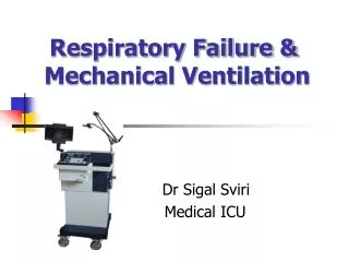
What Is Dyspnea? Dyspnea is a distressing sensation of difficult, labored, or unpleasant breathing.
What Is the Differential Diagnosis of Dyspnea? • There are a multiplicity of causes located in a variety of anatomic locations. • While the list of causes is nearly endless, 5 major causes account for ~94% of cases: • - Cardiac • - Respiratory • - Psychogenic/hyperventilation syndrome • - Deconditioning • - GERD
Classification of Dyspnea ClassPatient Symptoms Class I (Mild) No limitation of physical activity. Ordinary physical activity does not cause undue symptoms Class II (Mild) Slight limitation of physical activity. Comfortable at rest, ordinary physical activity results in symptoms. Class III (Moderate) Marked limitation of physical activity. Comfortable at rest, but less than ordinary activity causes symptoms. Class IV (Severe) Symptoms of cardiac insufficiency at rest. If any physical activity is undertaken, discomfort is increased.
Dyspnea: causes • Respiratory system • Obstruction: asthma, COPD, tumor • Pulmo Diseases: pneumonia, interstitial lung disease, • Pleura: pneumothorax, effusion • Diaphragma movement disorder: tense ascites • Cardiovascular system • Heart failure • Pulmo embolism
Features of left heart failure • Underlying diseases • Position related dyspnea • Crackles or rhonchi in both lungs • PND
Nocturnal paroxysmal dyspnea Characteristics • Awoken due to chest tightness or dyspnea • Forced sitting position or orthopnea • Tachycardia • crackles or rhonchi in both lungs • Pink frothy sputum
Mechanism • Vital capacity decreased in supine position • Returned blood volume increased pulmo edema Nocturnal paroxysmal dyspnea
Dyspnea: accompany signs (1) • Rhonchi • Asthma • Acute left heart failure (cardiac asthma) • Acute laryngeal edema • Chest pain • Infection • Pneumothorax • Pulmo embolism • Acute myocardial infarct
Dyspnea: accompany signs (2) • Fever • Infection • Cough and sputum • COPD • Infection • Left heart failure • Unconsciousness • CNS disorder • Uremia • diabetic ketoacidosis
What is the Value of History for Diagnosing the Etiology of Wheeze? • While wheezing is indicative of obstruction of airways, it is insensitive and nonspecific in diagnosing the specific location and cause of the obstruction. • Expiratory wheezing by history may be predictive of asthma no more than 35% of the time; past history of asthma, no more than 62%; monophonic expiratory wheezing by physical, no more than 43% of the time.
An Approach to the Diagnosis of Wheeze Be aware that: “All that wheezes is not asthma; All that wheezes is obstruction.”
Differential Diagnosis of Wheeze According to Anatomic Area UPPER AIRWAY OBSTRUCTION Extrathoracic Causes Intrathoracic Causes PNDS Tracheal stenosis Vocal cord dysfunction Goiter Syndrome Epiglottitis Malignancies Laryngeal edema Benign tumors Postextubation granuloma Anaphylaxis
Differential Diagnosis of Wheeze According to Anatomic Area LOWER AIRWAY OBSTRUCTION Asthma Bronchiolitis COPD Bronchiectasis Pulmonary edema Carcinoid syndrome Pulmonary embolism Parasitic infections Cystic fibrosis Lymphangitic carcinomatosis
Cyanosis • An excess of desaturated hemoglobin causes a blue coloration of the skin or mucosae.
Cyanosis: classification • Central (warm) • Deficient oxygenation • Right-to-left shunt • Peripheral (cold) • Reduced cardiac output • Local vasoconstriction • Mixed • Heart failure
Cyanosis: accompany signs • Dyspnea • Severe cardiac or respiratory disorder • Clubbing fingers • Congenital heart disease • Chronic respiratory disease
Suprasternal fossa Supraclavicular fossa Infraclavicular fossa Midclavicular line epigastric angle Anterior imaginary lines and landmarks
Posterior axillary line Anterior axillary line Midaxillary line Lateral imaginary lines
Suprascapular region Interscapular region Infrascapular region Scapular line Posterior midline Posterior imaginary lines and landmarks