Facial Injury Assessment
280 likes | 658 Vues
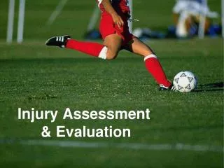
Facial Injury Assessment. Athletic Injury Assessment Chapter 17, p. 584. Ear Injuries: History p.585. Location of pain? External ear vs. internal ear Onset? Acute=trauma Gradual=infection Mechanism? Auricle trauma Slapping force to ear. Other symptoms: tinnitus dizziness possible.

Facial Injury Assessment
E N D
Presentation Transcript
Facial Injury Assessment • Athletic Injury Assessment • Chapter 17, p. 584
Ear Injuries: History p.585 • Location of pain? • External ear vs. internal ear • Onset? • Acute=trauma • Gradual=infection • Mechanism? • Auricle trauma • Slapping force to ear • Other symptoms: • tinnitus • dizziness possible
Inflammation of auricle (otitis externa) Auricle hematoma cauliflower ear fig. 17-11, p,. 592 Tympanic membrane eardrum WNL=shiny, translucent, smooth Ear Injuries: Observation p. 591 • Periauricular edema • Battle’s sign
Tympanic membrane eardrum WNL=shiny, translucent, smooth Ear Injuries: Observation p. 592
Ear Injuries: Palpation • Pericauricular area • r/o fx(mastoid process) • External ear (auricle)
Auricular hematoma--p. 598--Table 17-3 • Blunt trauma or shearing mechanism • “cauliflower ear” • pooling of blood between skin & auricular cartilage • may be drained/casted using flexible collodian
Tympanic membrane rupture--p.599 • Table 17-4, P. 599 • Mechanisms: • change in air pressure • direct blow to ear • inability to regulate internal pressure (infection) • injury from foreign object • Symptoms-- • intense pain • otoscopic exam may show drainage and perforation • hearing deficit
Otitis Externa--p.599 • “Swimmer’s Ear” • Cause: inadequate drying of the ear • Symptoms: • c/o pressure/pain in ear with possible itching • clear drainage possible • possible lymph node enlargement
Otitis media--p.465 • History: URI • Symptoms: • c/o pressure/pain in ear • pain worsens when earlobe is tugged • possible hearing deficit • otoscopic exam reveals opaque/bulging tympanic membrane
Clinical Anatomy: Teeth p.587 • Fig. 17-7, p. 587 • 32 permanent teeth • secured to gum by periodontal ligaments
Dental Injury Assessment--History • Location: usually easily pinpointed by athlete • Onset: usually acute • Mechanism: usually blunt force trauma (object/competitor)
Dental Injury Assessment--Inspectionp.594 • Assess any obvious fx • inner/outer surfaces • Assess bite/fit of teeth • Assess source of bleeding
Dental Injury Assessment--Palpationp.595 • Use caution! • Bites • Lacerations • Cross-contamination • Do not worsen the injury • Any abnormal looseness should be referred to physician
Dental Pathologiesp. 603 • Highest risk sport=Basketball • Mouthpieces decrease risk • custom • off-the-shelf/boil-and-bite • Tooth ID numbers: • fig. 17-22, p. 604
Tooth Fracture & Luxations--p. 604 • Luxations--fig. 17-24, p. 605 • Intruded/Extruded • may accompany pulp fx • angulation/rotation • self-evaluation • 90% success rate with reimplantation • Emergency Management- • Table17-9, p. 608 • Fractures: • Fig. 17-23, p.604 (I-IV) • Refer to dentist • Self-evaluated • Pain, temperature sensitivity, obvious deformity
Nose Injury Assessment--Historyp. 599 • Mechanism--usually direct trauma/impact • Insidious onset usually due to illness • May accompany other injuries (concussion) • Know PMH
Nasal Injuries--Observation • Alignment (fx?) • PMH? • Symmetry • Self-assessment • Bleeding (epistaxis) • “Raccoon eyes” • fig. 17-12, p. 593 • Saddle-nose deformity • fig.17-219, p. 600
Nasal Injuries--Palpationp. 594 • Facial bones • zygoma • orbit • frontal bone • Nasal bone • Nasal cartilage
Nasal Fracturep. 600 • Table 17-5, p. 600 • Deformity probable • Profuse bleeding • Crepitus with palpation possible • Breathing possibly obstructed
Epistaxis • Control bleeding for further evaluation • Methods: • pinch nostrils • ice packs • tilt head • Rolled gauze/tampon • antibiotic • topical decongestant • gauze under top lip
Clinical Anatomy: TMJp. 605 • Fig. 17-8, 606 • Synovial joint • temporal bone & mandible • Articular disk • attached to mandible • no attachment to capsule • TMJ glides & pivots
TMJ Assessment: History • Mechanisms: • trauma (subluxation) • teeth grinding/grating • dental malalignment • Onset • Acute, insidious, chronic • Clicking/popping • Headaches? • Stress levels?
TMJ Assessment: Observation • Malocclusion/deviation in bite • Obvious deformity possible • Difficulty speaking • Decreased AROM common (Fig. 17-18, p. 596) • Observe for excursion of TMJ motion • Grinding Teeth?
TMJ Palpation • Check for NML gliding/hinge action • Palpate for abnormal crepitus • Bilateral comparison • Palpate externally!
TMJ Testing • Knuckle Test • Box 17-2, p. 597 • (+) test= < 2 knuckles fitting in mouth
Throat Traumap. 600 • Blunt force trauma • Complications: • carotid sinus stimulation • respiratory interference from swelling • Aphasia • Cartilage displacement • Do not attempt to correct deviations • Refer to physician
LeFort Fracturesp. 602 • Midface fractures • 3 classes (I-III) • High impact force required (MVA) • Entire sections of face will displace • Uncommon in athletics • Fig. 17-21, p. 603