Download

1 / 136
1.36k likes | 1.79k Vues
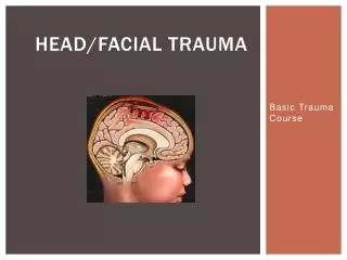
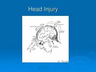
Head and Facial Injury. Scott Marquis, MD. Overview. Head injury What to look for Appropriate management Facial injury Review. Head and brain trauma. ~ 1,500,000 head injuries annually ~ 230,000 hospitalized and survive ~ 50,000 deaths 1/3 all injury-related deaths Severity 75% mild

E N D
Head and Facial Injury Scott Marquis, MD
Overview • Head injury • What to look for • Appropriate management • Facial injury • Review
Head and brain trauma • ~ 1,500,000 head injuries annually • ~ 230,000 hospitalized and survive • ~ 50,000 deaths • 1/3 all injury-related deaths • Severity • 75% mild • 10% moderate • 10% severe (35% mortality, 5% c-spine fx) • 80,000-90,000 significant long-term disability
Head & brain trauma • Risk Groups • Highest: Males 15-24 yrs of age • Very young children: 6 mos to 2 yrs of age • Young school age children • Elderly >75 yrs
Head injury • Broad and Inclusive Term • Traumatic insult to the head that may result in injury to soft tissue, bony structures, and/or brain injury • Blunt Trauma • Penetrating Trauma
Brain injury • “A traumatic insult to the brain capable of producing physical, intellectual, emotional, social and vocational changes” • Three broad categories • Focal injury • Cerebral contusion • Intracranial hemorrhage • Epidural hemorrhage • Subarachnoid hemorrhage • Diffuse Axonal Injury • Concussion
Mechanisms of head injury • Motor vehicle crashes, MVC • Most common cause of head trauma • Most common cause of subdural hematoma • Sports injuries • Falls • Common in elderly and in presence of alcohol • Associated with subdural hematomas • Penetrating trauma • Missiles more common than sharp projectiles
Categories of injury • Coup injury • Directly posterior to point of impact • More common when front of head struck • Contrecoup injury • Directly opposite the point of impact • More common when back of head struck
Categories of injury • Diffuse axonal injury (DAI) • Shearing, tearing or stretching of nerve fibers • More common with vehicle occupant and pedestrian • Focal injury • Limited and identifiable site of injury
Causes of brain injury • Direct (primary) causes • Impact • Mechanical disruption of cells • Vascular permeability or disruption • Indirect (secondary or tertiary) causes • Secondary • Edema, hemorrhage, infection, inadequate perfusion, tissue hypoxia, pressure • Tertiary • Apnea, hypotension, pulmonary resistance, ECG changes
Brain anatomy • Occupies 80% of intracranial space • Divisions • Cerebrum • Cerebellum • Brain Stem
Brain anatomy • Cerebral spinal fluid, CSF • Clear, colorless • Circulates throughout brain and spinal cord • Cushions and protects • Ventricles • Center of brain • Secrete CSF by filtering blood • Forms blood-brain barrier
Brain anatomy • Blood Supply • Vertebral arteries • Supply posterior brain (cerebellum and brain stem) • Carotid arteries • Most of cerebrum
Brain anatomy • Meninges • Dura mater: tough outer layer, separates cerebellum from cerebral structures, landmark for lesions • Arachnoid: web-like, venous vessels that reabsorb CSF • Pia mater: directly attached to brain tissue
Scalp lacerations • Scalp laceration or avulsion • Most common injury • Vascularity = diffuse bleeding • Generally does not cause hypovolemia in adults • Can produce hypovolemia in children
Scalp anatomy • Scalp • S: skin • C: connective tissue • A: aponeurosis (galea) • L: loose areolar tissue • P: pericranium • Scalp very vascular • major blood loss • watch kids and adults with prolonged extrication
Skull fracture • Present in 60% of pts with severe head injury • Types: • Linear: usually incidental finding on CT • Depressed: mechanism is usually intense blow to scalp with object of small surface area. Surgical repair needed if depressed more than 5mm
Skull fracture • Types • Basilar: blow to temporal (most often), parietal, occipital area • Signs • Hemotympanum or bloody ear discharge • Rhinorrhea or otorrhea • Battle’s sign • Racoon’s eyes • Cranial nerve palsies
Closed head injuries • Focal • Contusion • Epidural hematoma • Subdural hematoma • Intracerebral • Diffuse (most common type of head injury) • Mild concussion • Classic concussion • Diffuse Axonal Injury (DAI)
Blood between skull and dura Usually arterial tear Middle meningeal artery Causes increased ICP Epidural hematoma
Epidural hematoma • Unconsciousness followed by lucid interval • Rapid deterioration • Decreased LOC, headache, nausea, vomiting • Hemiparesis, hemiplegia • Unequal pupils (dilated on side of clot) • Increase BP, decreased pulse (Cushing’s reflex)
Between dura mater and arachnoid More common Usually venous Bridging veins between cortex and dura Causes increased intracranial pressure Subdural Hematoma
Subdural hematoma • Slower onset • Increased ICP • Headache, decreased LOC, unequal pupils • Increased BP, decreased pulse • Hemiparesis, hemiplegia
Intracerebral hematoma • Usually due to laceration of brain • Bleeding into cerebral substance • Associated with other injuries • DAI • Neuro deficits depend on region involved and size • Repetitive with frontal lobe • Increased ICP
Concussion • Transient loss of consciousness • Retrograde amnesia, confusion • Resolves spontaneously without deficit • Usually due to blunt head trauma
Diffuse axonal injury • Tearing or shearing of nerve fibers at time of impact • Rapid acceleration-deceleration injury (MVA) • Functional or physiologic dysfunction • Not gross anatomic abnormality • Most common CT finding after severe head trauma
Diffuse axonal injury • Prolonged post-traumatic coma not due to mass lesion or ischemic insults • Coma begins at time of trauma • Usually evidence of decorticate or decerebrate posturing, autonomic dysfunction (HTN, fever)
Penetrating head injury • Severity depends on • Energy of missile • Path • Amount of scatter of bone and metal fragments • Presence of mass lesion • Accompanied by • Severe face and neck injuries • Significant blood loss • Difficult airway • Spinal instability
What the brain needs • High metabolic rate • Consumes 20% of body’s oxygen • Largest user of glucose • Requires thiamine • Can not store nutrients
More on brain workings • Perfusion • Cerebral blood flow (CBF) • Dependent upon CPP • Flow requires pressure gradient • Cerebral perfusion pressure (CPP) • Pressure moving the blood through the cranium • Autoregulation allows BP change to maintain CPP • CPP = mean arterial pressure (MAP) - intracranial pressure (ICP)
More on brain workings • Perfusion • Mean Arterial Pressure (MAP) • Largely dependent on cerebral vascular resistance (CVR) since diastolic is main component • Blood volume and myocardial contractility • MAP = diastolic + 1/3 pulse pressure • Usually require MAP of at least 60 mm Hg to perfuse brain
More on brain workings • Perfusion • Intracranial pressure (ICP) • Edema, hemorrhage • ICP usually 10-15 mm Hg • Cerebral perfusion pressure CPP = MAP - ICP
What goes wrong in head injury • As ICP and approaches MAP, cerebral blood flow • Results in CPP • Compensatory mechanisms attempt to MAP • As CPP , cerebral vasodilation occurs to blood volume • This leads to further ICP, CPP and so on