CASE 3
CASE 3. Reyes, Carmen; Reyes, Jenilene ; Reyes, Lourdes; Rivera, Laila ; Rivere , Djeaune ; Robosa , Dean Antonio; Rodas , Francis; Rodriquez, Reine ; Rogelio, Graciella ; Roque , Marianne; Ruanto , Maria Theresa. Outline of Presentaion.


CASE 3
E N D
Presentation Transcript
CASE 3 Reyes, Carmen; Reyes, Jenilene; Reyes, Lourdes; Rivera, Laila; Rivere, Djeaune; Robosa, Dean Antonio; Rodas, Francis; Rodriquez, Reine; Rogelio, Graciella; Roque, Marianne; Ruanto, Maria Theresa
Outline of Presentaion • To present a case of a 24F presenting with shortness of breath • To present an approach and an algorithm in the diagnosis of a patient with shortness of breath • To present the differential diagnosis and clinical impression of the patient • To discuss the chest x-ray findings and correlate it with the PE examination findings • To discuss the roles of other imaging modalities 2
Outline of Presentaion • To present a case of a 24F presenting with shortness of breath • To present an approach and an algorithm in the diagnosis of a patient with shortness of breath • To present the differential diagnosis and clinical impression of the patient • To discuss the chest x-ray findings and correlate it with the PE examination findings • To discuss the roles of other imaging modalities 3
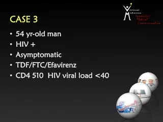
CASE PRESENTATION • R.F. • 24 y/o • Female • CHIEF COMPLAINT: Shortness of breath 4
HISTORY OF PRESENT ILLNESS 5 ADMISSION
REVIEW OF SYSTEMS • Poor appetite • No headache/blurring of vision • No cough/colds • Occasional chest pain • No abdominal pain/no vomiting • No joint pains 6
PAST MEDICAL HISTORY • No previous hospitalizations • (+) episodes of sore throat and fever as a child • No hypertension • No diabetes • No surgeries 7
FAMILY HISTORY • (-) Heart disease • (-) Diabetes • (-) Asthma/Allergies 8
PERSONAL/SOCIAL HISTORY • Non-smoker • Non-alcoholic beverage drinker 9
PHYSICAL EXAMINATION FINDINGS • Conscious, coherent, ambulatory • BP: 120/80 • HR: 70 bpm • RR: 20’s • Warm moist skin ,no dermatoses • HEART: apex beat 5th LICS, MCL (+) accentuated S1 (+) diastolic murmur • LUNGS: symmetric chest expansion no retractions (+) occasional wheeze • No cyanosis/edema 10
MISSING DATA • General Data • Address, occupation, civil status, religion • HPI • Type of vitamins taken when consult was done • Other possible associated signs and symptoms • PE findings • Specific RR • Temp • BMI • JVP • Personal and Social History • Type of diet, exercise • Occupation (type, workload) • Environmental History • Area of residence and associated living conditions 11
SALIENT FEATURES • 24 F • BP: 120/80 • HR: 70 bpm • RR: 20s • Progressive shortness of breath • Symmetrical chest expansion • (-) Retractions • (+) Wheeze • Apex beat: 5th LICS, MCL • (+) Accentuated S1 • (+) Diastolic murmur 12
Outline of Presentaion • To present a case of a 24F presenting with shortness of breath • To present an approach and an algorithm in the diagnosis of a patient with shortness of breath • To present the differential diagnosis and clinical impression of the patient • To discuss the chest x-ray findings and correlate it with the PE examination findings • To discuss the roles of other imaging modalities 13
Dyspnea • a subjective experience of breathing discomfort that consists of qualitatively distinct sensations that vary in intensity • derives from interactions among multiple physiological, psychological, social, and environmental factors, and may induce secondary physiological and behavioural responses 17
Association of Qualitative Descriptors and Pathophysiologic Mechanisms of Shortness of Breath 19
Chest Pain • discomfort or pain anywhere along the front of your body between your neck and upper abdomen • Can be due to cardiopulmonary problems, chest wall problems, GI, psychological 21
Apex beat displacement • Patient AB: 5th LICS, MCL • Lateral and/or inferior displacement of the apex beat usually indicates cardiomegaly. • May also be displaced by other conditions: • Pleural or pulmonary diseases • Deformities of the chest wall or the thoracic vertebra 23
(+) accentuated S1 • Mitral valve snaps shut more vigorously, producing a louder S1 • Blood velocity is increased-> anemia, fever, hyperthyroidism, anxiety, and during exercise • Mitral valve is stenotic 24
(+) diastolic murmur • Heard with bell at apex, patient in left lateral decubitus position • Findings on examination • Low-frequency diastolic rumble, more intense in early and late diastole, does not radiate; systole usually quiet; palpable thrill at apex in late diastole common; S1 increased and palpable at left sternalborder • Description • Narrowed valve restricts forward flow; forceful ejection into the ventricle • Often occurs with mitral regurgitation caused by rheumatic heart fever or cardiac infection 26
Lung Findings Occasional wheeze • Musical respiratory sounds thaat may be audible both to the patient and to others • Suggests partial airway obstruction from secretions, tissue inflammation, or a foreign body. 27
Outline of Presentaion • To present a case of a 24F presenting with shortness of breath • To present an approach and an algorithm in the diagnosis of a patient with shortness of breath • To present the differential diagnosis and clinical impression of the patient • To discuss the chest x-ray findings and correlate it with the PE examination findings • To discuss the roles of other imaging modalities 28
Outline of Presentaion • To present a case of a 24F presenting with shortness of breath • To present an approach and an algorithm in the diagnosis of a patient with shortness of breath • To present the differential diagnosis and clinical impression of the patient • To discuss the chest x-ray findings and correlate it with the PE examination findings • To discuss the roles of other imaging modalities 29
Trachea Patient’s PA CXR Normal PA CXR (-) tracheal deviation (-) flattening of the R&L hemidiaphragm (-) pulmonary congestion (-) blunting of the costophrenic angle (-) bone deformities (-) pulmonary infiltrates
Patient’s PA CXR Normal PA CXR 32 (+) heart enlargement Slight straightening of the L cardiac border
Normal PA CXR Normal location of the apex: 5th ICS, MCL 33
Patient’s PA CXR The patient’s apex is located on the 7th ICS MCL – DOWNWARD DISPLACEMENT OF THE APEX 34
Cardio-thoracic Ratio Normal PA CXR Patient’s PA CXR
Which chamber/s is/are enlarged? Normal LVE LAE & LVE (in long-standing MS) LAE 1 – R brachiocephalic vessels 2 – Ascending aorta and superimposed SVC 3 – R atrium 5 – L brachiocephalic vessels 6 – Aortic arch 7 – Pulmonary trunk 8 – L atrial appendage 9 – L ventricle 36 Squire’s Fundamentals of Radiology, 6th ed.
Normal PA CXR Patient’s PA CXR Left Atrial Enlargement Prominent L atrial appendage 37
Normal PA CXR Patient’s PA CXR Carina not appreciated (cannot be measured for widening) 38
Normal PA CXR Patient’s PA CXR No double density along the R cardiac border 39
Possible L Ventricular Enlargement Normal PA CXR Patient’s PA CXR Downward dipping of the left heart
Possible L Ventricular Enlargement Normal PA CXR Patient’s PA CXR LV outflow tract
Possible R Ventricular Enlargement Normal PA CXR Patient’s PA CXR Rounding of the cardiac apex
Normal Lateral CXR Patient’s Lateral CXR Trachea Trachea Esophagus Esophagus Heart Heart 43
Left atrial enlargement Esophagus Esophagus Retrocardiac free space Retrocardiac free space 44
Left venticular enlargement LV outflow tract LV outflow tract Left cardiac border Left cardiac border 45
Left venticular enlargement Posterior margin of the IVC Obliterated posterior margin of the IVC Convex posterior heart border 46
Left venticular enlargement Hoffman Rigler sign 47
Right ventricular enlargement Retrosternal space Retrosternal space 1/3 2/3 48
Outline of Presentaion • To present a case of a 24F presenting with shortness of breath • To present an approach and an algorithm in the diagnosis of a patient with shortness of breath • To present the differential diagnosis and clinical impression of the patient • To discuss the chest x-ray findings and correlate it with the PE examination findings • To discuss the roles of other imaging modalities 49