Understanding Thrombin Inhibition: Structure, Function, and Classification
Explore thrombin inhibition through structural analysis, key sites, and inhibitor classification using bioinformatics tools and databases. Learn about the significance of inhibiting thrombin in preventing blood clot formation.

Understanding Thrombin Inhibition: Structure, Function, and Classification
E N D
Presentation Transcript
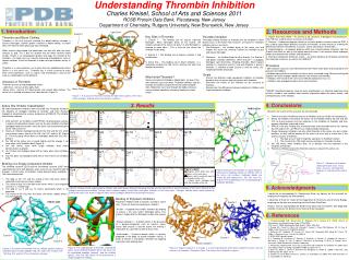
Understanding Thrombin Inhibition Charles Kreisel, School of Arts and Sciences 2011 RCSB Protein Data Bank, Piscataway, New Jersey Department of Chemistry, Rutgers University, New Brunswick, New Jersey 1. Protein Data Bank (PDB) – An archive of 3D structures of biological macromolecules. The PDB was used to find all structures of thrombin. 2. Chimera – A computer program which can visualize protein structures from the PDB. It was used to superimpose different structures of thrombin, to make movies of showing the differences between the different structures, and to take pictures of thrombin 4. 3. Ligand Explorer – A computer program which can visualize protein structures from the Protein Data Bank as well as the ligands bound to them. It can analyze where hydrogen bonds form between the inhibitor and the protein 5. 4. Chemical Component Dictionary (CCD) 6– An online database of all ligands containing their chemical structures as well as their SMILES*. Procedure –364 thrombin structures were identified from the Protein Data Bank and analyzed for the presence of inhibitors. –Inhibitors bound to these thrombin structures were visualized using Chimera and Ligand Explorer to find all hydrogen bonds between the inhibitor and thrombin. – Inhibitors were found in the CCD and a SMILES comparison was run. – Inhibitors were grouped based on these classifications. *SMILES (simplified molecular input line entry specification) is a chemical code consisting of letter, numbers, and symbols which contain information about the atoms, bonds, and stereochemistry of the inhibitor. 1. Introduction 2. Resources and Methods Light Chain Exosite 1 Key Sites in Thrombin 1. Exosite 1 – The binding site for natural substrate fibrinogen. When fibrinogen binds to the exosite, the negative charge of the active site pulls it in and fibrinogen is cleaved to make fibrin 2. This is also the site where the inhibitor hirudin binds. 2. Exosite 2 – The binding site of heparin cofactor II and antithrombin 2. 3. Active Site – The binding site of direct inhibitors. It is marked by residues Ser 195, His 57, and Asp 102 and is the location for catalysis. Why Inhibit Thrombin? Venous and arterial thrombosis (blood clots) are one of the most common causes of death. Thrombin inhibitors stop the function of thrombin and prevent the formation of blood clots. Blood clots can travel through the body and cause several medical conditions ultimately leading to a heart attack 1. • Thrombin Inhibition • The blood clotting function of thrombin can be inhibited in three distinct ways: at the Active Site, Exosite I, or Exosite II. Thrombin inhibitors come in two forms. • Non-Polymeric - the inhibitor binds to the active site and morphs the active site, making it difficult for thrombin to pull and cleave fibrinogen • Polymeric - the inhibitor binds to either Exosite 1 or Exosite 2 and blocks thrombin differently1. When Exosite 1 is plugged, fibrinogen can’t bind there, inhibiting thrombin. When heparin cofactor II is bound to exosite 2 and anti-thrombin is present, thrombin is inhibited at both Exosite 2 and the active site, morphing thrombin drastically. PDB ID 1SGI Thrombin and Blood Clotting Thrombin is the final enzyme involved the blood clotting cascade. It cleaves fibrinogen, another protein involved in blood clotting, to create fibrin, the material which physically clots the blood. When leeches draw blood, the blood does not clot until after the leech releases its grip. This is due to a protein that the leech secretes called hirudin. Hirudin is an anticoagulant protein and stops the function of the thrombin1. Our body also has naturally occurring anticoagulants such as heparin cofactor II and anti-thrombin in order to keep thrombin activity in check 1. Thrombin is a serine protease, an enzyme that cuts peptide bonds where Serine is in the active site 2. Thrombin has a similar structure to many other serine proteases, such as trypsin and chymotrypsin, and can be used as a prototypical serine protease. Structure of Thrombin Thrombin consists of two polypeptide chains: -Light chain - consists of one alpha helix -Heavy chain - consists of 2 beta barrels and several alpha helices. The active site is located in between the 2 beta barrels (Figure 1). • Goals • Classify the different single component inhibitors of thrombin based on their structures and interactions with thrombin. • Determine the effects of the inhibitors on the structure of thrombin. • Demonstrate the differences between polymeric inhibitors and single component inhibitors. Exosite 2 Heavy Chain Figure 1: A structure of thrombin showing the light chain (green), the heavy chain (orange), and the active site residues (yellow) 3. 4. Conclusions 3. Results • Active Site Inhibitor Classification • 58 single component inhibitors were classified into 19 groups based on the locations of hydrogen bonding to thrombin and their chemical fingerprints, determined by using a comparison of SMILES. The inhibitor classification showed: • 0G6 and 0G7 are inhibitors called PPACK (D-phenylalanyl-L-prolyl-L-arginine chloromethyl ketone) and are the only inhibitors that both hydrogen bond with residues near the active site and covalently bind to Ser 195 and His 57. • Nearly all inhibitors hydrogen bond with Gly 216 and Ser 214 which are residues across from the Ser 195, Asp 102, and His 57 (Figure 5). The only group which does not hydrogen bond with Gly 216 is group 3. • Ser 195 of the active site is turned slightly and the change is not large when small inhibitors bind (Figure 3). • Ser 195 moves more when larger inhibitors bind (video demonstration available). • His 57 does not hydrogen bond with or move when many inhibitors bind. • Asp 102 does not hydrogen bond with or move when any inhibitors bind • Binding of a Single Component Inhibitor • The inhibited structure 2ZC9 and the uninhibited structure 2UUF were superimposed and a path between each structure was traced to look for changes in other areas of thrombin (video demonstration available) 7. The traces determined: • The loop at Thr 147 and the strand at Glu246 move which is expected since they are always mobile. • The strand at Asp 14 in the light chain moves which is also expected since this is a mobile loop. • The loop at Lys 9 and Lys 10 moves significantly which is not expected. • The strand at Glu 192 near the active site moves slightly which is also not expected. PDB ID 2ZC9 • Based on the results of this research, we can conclude: • There are at least 19 different classes of inhibitors that can inhibit serine proteases. • Nearly all inhibitors that bind to the active site of thrombin bind to Gly 216 and Ser 214, so these residues must be important in the inhibition of thrombin and their purpose should be examined further. • PPACK is the only inhibitor that covalently binds to the thrombin active site, moving Ser 195 and His 57, so PPACK must inhibit thrombin differently. • Single Component inhibitors not only affect thrombin at the active site, but in other locations as well and the position of the active site must play a crucial role in the overall structure of thrombin. • 4.1 The loop at Lys 9 and 10 might be significant in thrombin function. • 4.2 The strand at Glu 192 might also be significant in thrombin function. • Ser 195 moves when inhibitors bind, so its position must be important in the inhibition of thrombin. • Polymeric inhibitors bind to the thrombin exosites instead of the active site. Gr9 0G6 0G7 Gr1 22U 37U 51U 53U 19U 176 177 16U 21U Gr5 34P 3SP QWE 00N 00Q Gr13 2TS Gr17 BM2 BM9 0G6 3SP 22U 2TS BM2 Ser 195 Ser 214 Asp 102 00L 24U 0KV GAH 0E7 Gr2 32U 45U 46U 50U 29U 27U 24U 31U 33U 49U Gr6 0IT 0IV 0KV 0ZG 0ZI CCR Gr10 GAH Gr14 00L 00K Gr18 0E7 His 57 ALZ 10U MID 1Z0 MDL Gr7 MID MIT Gr15 AZL ALZ Gr3 10U 11U 12U 13U Gr11 MDL Gr19 1Z0 Light Chain Exosite 1 -Hirudin -Fibrinogen Figure 7: A diagram of thrombin showing the light chain, heavy chain, exosites, and important residues in and around the active site. Ser 195 Ser 214 RA4 165 Gr4 RA4 RA8 Gr8 162 163 162 Gr12 165 Gr16 T19 T42 Figure Legend Regions which differ from other inhibitors in the same class. Atoms which covalently bond to the thrombin active site. Atoms which hydrogen bond with the thrombin active site. T42 Figure 5:Structure of thrombin in Ligand Explorer showing the hydrogen bonds of inhibitor 22U to the active site residues. Hydrogen bonds are shown by pink dotted lines. Ser 195, Asp 102, and His 57 are labeled but are not participating in hydrogen bonding 8. Exosite 2 -Antithrombin -Heparin Cofactor II His 57 Gly 216 Asp 102 Active Site -Single Component inhibitors Heavy Chain 5. Acknowledgments Figure 4: Images of one representative inhibitor from each group. Beneath the group number are the ligand IDs of all the inhibitors in that class. Several of the groups have only one inhibitor. Orange circles indicate regions which differ from other inhibitors in the same class. Triangles indicate atoms which covalently bind to the active site. Arrows indicate atoms which hydrogen bond with the thrombin residues. I would like to acknowledge Dr. Shuchismita Dutta, my advisor for this research for helping me throughout the whole project. I would like to thank Dr. Taylor of the Department of Chemistry and Chemical Biology for giving me the idea of researching with the Protein Data Bank. Finally I want to acknowledge the Protein Data Bank and researchers who deposited thrombin structures for giving me the material to do my project. Binding of Polymeric Inhibitors Polymeric inhibitors bind to exosites instead of active sites. There are three main polymeric inhibitors. -Hirudin – A peptide that inhibits thrombin by binding to exosite I, the area where fibrinogen binds. This makes it impossible for fibrinogen to dock into exosite 1. -Heparin cofactor II – A protein which, in the presence of a glycosaminoglycan such as heparin, spans across both exosite 2 and the active site making it impossible for substrates to bind to those sites. -Antithrombin – A plasma protein which is naturally produced in the body to control thrombin activity. It binds to exosite 2 and blocks thrombin by stopping substrates from binding there. PDB ID 1JMO PDB ID 2ZC9 Exosite 2 PDB ID 2ZC9 6. References 1. Tanaka-Azevedo A.M, Morais-Zani K, Torquato R.J.S, Tanaka A.S. (2010) Journal of Biomedicine and Biotechnology. August 24;641025. 2.Di Cera E. (2008) Mol Aspects Med. August; 29(4): 203–254. 3. Pineda AO, Carrell CJ, Bush LA, Prasad S, Caccia S, Chen ZW, Mathews FS, Di Cera E. (2004) J Biol Chem.Jul 23;279(30):31842-53. 4. Pettersen EF, Goddard TD, Huang CC, Couch GS, Greenblatt DM, Meng EC, Ferrin TE. (2004) /J Comput Chem./ Oct;25(13):1605-12. 5. RCSB PDB Ligand Explorer 6. J. Callaway, M. Cummings, B. Deroski, P. Esposito, A. Forman, P. Langdon, M. Libeson, J. McCarthy, J. Sikora, D. Xue, E. Abola, F. Bernstein, N. Manning, R. Shea, D. Stampf, and J. Sussman (1996) Brookhaven National Laboratory. 7. Ahmed HU, Blakeley MP, Cianci M, Cruickshank DW, Hubbard JA, Helliwell JR. (2007) Acta Crystallogr D Biol Crystallogr. Aug;63(Pt 8):906-22. Epub Jul 17. 8. Baum B, Muley L, Heine A, Smolinski M, Hangauer D, Klebe G. (2009) J Mol Biol. Aug 21;391(3):552-64. Epub Jun 9. 9. Baglin TP, Carrell RW, Church FC, Esmon CT, Huntington JA. (2002) Proc Natl Acad Sci U S A. Aug 20;99(17):11079-84. Exosite 1 Exosite 1 Thrombin Exosite 2 Heparin cofactor II Figure 3: The superposition of thrombin showing the active site of inhibited thrombin (blue), the active site of uninhibited thrombin (orange), and the inhibitor (green). Only Ser 195 moves in the presence of a single component inhibitor 8. Figure 6: Heparin cofactor II, in orange, is a very large cofactor which binds to both the active site and exosite 1 of thrombin. Thrombin is blue. The active site of thrombin is green 9. Figure 2: A structure of thrombin with an inhibitor (green) showing thrombin (blue), hirudin peptide (red), the active siteresidues Ser 195, Asp 102, and His 57 are showing in orange 8.