Articulations Chapter 9
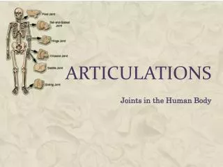
Articulations Chapter 9. Biology 210 Instructor: John McGill Original PowerPoint: Jack Bagwell Supplemental Notes: Beth Wyatt Last updated: July 15, 2014. INTRODUCTION TO ARTICULATIONS. DEFINITION Articulations (joints) are the junctions between bones. FUNCTION

Articulations Chapter 9
E N D
Presentation Transcript
ArticulationsChapter 9 Biology 210 Instructor: John McGill Original PowerPoint: Jack Bagwell Supplemental Notes: Beth Wyatt Last updated: July 15, 2014
INTRODUCTION TO ARTICULATIONS • DEFINITION • Articulations (joints) are the junctions between bones. • FUNCTION • They bind the various parts of the skeletal system together. • They permit bone growth and enable body parts to move in response to skeletal muscle contractions.
CLASSIFICATION-SUMMARY • Joints vary greatly in structure and function. • They can be classified by the types of tissues that bind the bones together at junctions • Synarthroses-fibrous • Amphiarthroses-cartilaginous • Diarthroses-synovial
CLASSIFICATION OF JOINTS: Structural • Based on Design, There Are 3 Types of Joints • FIBROUS JOINTS • Fibrous Tissue Located B/T Bones • CARTILAGINOUS JOINTS • Cartilage Located B/T Bones • SYNOVIAL JOINTS • Fluid-Filled Space Located B/T Bones
CLASSIFICATION OF JOINTS: Functional • Based on Degree of Movement Permitted, There Are 3 Types of Joints • Synarthroses (fibrous) • no movement (or very limited movement), suture • bones at these joints are fastened tightly by a layer of fibrous connective tissue • Amphiarthroses (cartilagenous) • cartilaginous joints • slightly moveable, symphysis pubis • bones at these joints are connected by hyaline cartilage • Diarthrosis (synovial) • freely moveable, shoulder joint • 7 major components of these joints
Synarthroses (Fibrous joints) • Syndesmoses • Sutures • Gomphoses
Synarthroses (Fibrous joints) • Syndesmoses • Bones are bound together by ligaments • Limited movement is possible • Ex: Joint between distal end of radius and ulna
Synarthroses (Fibrous joints) • Sutures • Occur only between the flat bones of the skull. • United by a connective tissue called a sutural ligament; eventually this ligament becomes ossified. • Joints between skull bones
Synarthroses (Fibrous joints) • Gomphoses • The union of a cone-shaped bony process in a bony socket. • Ex: a tooth is fastened to the jawbone by a peridontal ligament.
Amphiarthroses (Cartilaginous Joints) • Synchondroses • Sympheses
Amphiarthroses (Cartilaginous Joints) • Synchondroses • United by bands of hyaline cartilage. • Many of these joints are temporary structures that disappear as a result of the growth process • Examples • Epiphyseal plate between the epiphysis and the diaphysis • costal cartilage (ribs articulate with sternum)
Amphiarthroses (Cartilaginous Joints) • Sympheses • Joint in which a pad or disk of fibrocartilage connects two bones • Most are located in the midline of the body. • Example: • symphysis pubis • intervertebral disks
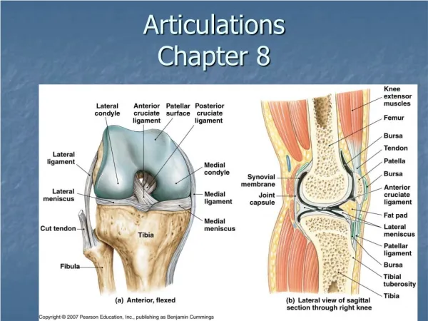
Structure of the Synovial (Diarthroses) Joint: Elbow, Knee, Shoulder, Hip, etc… • Joint capsule • sleeve like extension of the periosteum • forms a covering around the ends of the bones and binds the bones together. • Synovial membrane • moist membrane which lines the inner surface of the joint capsule • secretes synovial fluid • Articular cartilage • covers and cushions the articulating surfaces of the bones
Structure of the Synovial (Diarthroses) Joint: • Joint cavity • small space between the articulating surface of the 2 bones of the joint
Structure of the Synovial (Diarthroses) Joint: Elbow, Knee, Shoulder, Hip, etc… • Mensci (articular disk) • pads of cartilage between the articulating ends of bone
Structure of the Synovial (Diarthroses) Joint: Elbow, Knee, Shoulder, Hip, etc… • Ligaments • helps hold the articular cartilages together
Structure of the Synovial (Diarthroses) Joint: Elbow, Knee, Shoulder, Hip, etc… • Bursae • pillow-like structure formed from the synovial membrane
TYPES AND RANGE OF MOVEMENT AT SYNOVIAL/DIARTHROSES • Angular movements • Flexion • decreases the angle between two bones • “bending” a body part • Extension • increases the angle between two bones • “straightening” a joint
ANGULAR DIARTHROTIC MOVEMENT • Angular movements • PLANTAR FLEXION • Straightening the Foot • Downward (Points Toes Downward) • DORSIFLEXION • Bending the Foot Upward
ANGULAR DIARTHROTIC MOVEMENT Angular movements continued… • Abduction • move a body part away from the midline • Adduction • moves body part toward the midline
CIRCULAR DIARTHROTIC MOVEMENT • CIRCULAR MOVEMENTS • ROTATION • Bone Pivots Around a Fixed Point • CIRCUMDUCTION • Moves a Body Part so That Its Distal End Describes a Circle
CIRCULAR DIARTHROTIC MOVEMENT • CIRCULAR MOVEMENTS • SUPINATION • Moves the Forearm so as to Turn the Palm Up • PRONATION • Moves the Forearm so as to Turn the Palm Down
GLIDING DIARTHROTIC MOVEMENT • GLIDING MOVEMENTS • Sliding Between Flat Surfaces • Carpals & tarsals • Articular facets
GLIDING DIARTHROTIC MOVEMENT • SPECIAL MOVEMENTS • INVERSION • Turns the Sole of the Foot Inward • EVERSION • Turns the Sole of the Foot Outward
SPECIAL DIARTHROTIC MOVEMENT • SPECIAL MOVEMENTS continued… • PROTRACTION • Moves a Body Part Forward • Sticking out jaw • RETRACTION • Moves a Body Part Backward
SPECIAL DIARTHROTIC MOVEMENT • SPECIAL MOVEMENTS continued… • ELEVATION • Raises a Body Part • Closing one’s mouth • DEPRESSION • Lowers a Body Part • Opposite of elevation
TYPES OF SYNOVIAL/DIARTHROSES (Moveable) • Uniaxial joints • Biaxial joints • Multiaxial joints
Uniaxial joints-Hinge • Permit movement around one axis and in one plane. • Hinge joints • the convex surface of one bone fits into the concave surface of another bone. • Elbows, phalanges, knee • Flexion & Extension
Uniaxial joints-Pivot • Permit movement around one axis and in one plane. • Pivot joints • the cylindrical surface of one bone rotates within a ring formed from the fibrous tissue of a ligament. • Neck turning (1st and 2nd cervical vertebrae) • Rotation
Biaxial Joints-Saddle • Permit movement around two perpendicular axes in 2 perpendicular planes. • Saddle joints • formed between bones whose articulating surfaces have both convex and concave regions. • Thumb joint between first metacarpal and carpal bone • Flexion, extension, abduction, adduction
Biaxial Joints-Condyloid • Permit movement around two perpendicular axes in 2 perpendicular planes. • Condyloid (ellipsoidal) joints • a condyle fits into an elliptical socket. • Joint between radius and carpal bones • Flexion, extension, abduction, adduction
Multiaxial joint-Gliding • Joint that permits movement around 3 or more planes • Gliding joints • articulating surfaces are nearly flat or slightly curved • these joints only sliding (back and forth) motion (least movable) • Processes between vertebrae (articular facets) • gliding
Multiaxial joint-Ball and Socket • Joint that permits movement around 3 or more planes • Ball and socket joints • consists of a bone with a slightly egg-shaped head that articulates with the cup-shaped cavity of another bone • allows for the broadest range of movements. • Shoulder and hip • Flexion, extension, abduction, adduction, rotation, circumduction
REPRESENTATIVE SYNOVIAL JOINTS • SHOULDER JOINT • HIP JOINT • KNEE JOINT
SHOULDER JOINT • The Most Moveable Diarthrosis • Reason: Glenoid Cavity (Scapula) Shallow, Head of Humerus Doesn’t Fit Deep
HIP JOINT • The Most Stable Diarthrosis • Reason: Acetabulum (Os Coxa) Deep, Head of Femur Fits Deep
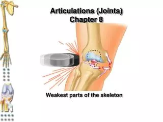
KNEE JOINT • The Major Weight Bearing Diarthrosis • The Most Frequently Injured Diarthrosis • Reasons • Fit Between Femur and Tibia (Condyles) Unstable • Little Muscle Over Knee Joint
Anatomy of the Knee http://www.aclsolutions.com/images/Seif_what is ACL.jpg
Diseases Normal Femur Osteoporotic Head
Resources • http://www.innerbody.com/image/skelfov.html • Publisher site: http://evolve.elsevier.com/ProductPage?product=0323016286
Case Study • http://www.recoverymedicine.com/rheumatoid_arthritis.htm • http://www.recoverymedicine.com/tendinitis_bursitis.htm