Download
1 / 52
590 likes | 864 Vues
PRESENTED BY: Dr. Areej Al- jabaly. ECG In Miscellaneous Conditions . A: Drugs Effects B: Electrolyte C : Diseases D: Normal Variants . Digoxin : Therapeutic Effect * ST segment depression ( reversed tick ). A: Drugs Effects .

E N D
PRESENTED BY: Dr. Areej Al-jabaly ECG In Miscellaneous Conditions
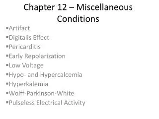
A: Drugs Effects B: Electrolyte C : Diseases D: Normal Variants
Digoxin : Therapeutic Effect * ST segment depression ( reversed tick ) A: Drugs Effects
Toxic Effect : Any type of arrhythmia especiall ventricular octopi
Quinidine:and related drugs like ( procinamide , Disopyramide , phenothiazine, Tricyclic, Antidepressant, Amiodarone) * P wave widening * QRS widening * Prolonged QT interval ( longer than half of the RR interval)
* Increase U wav amplitude * ST segment depression * Increase U wav amplitude
B: Electrolyte : Hyperkalemia : 1- Mild to moderate hyperkalemia (5 -7 mEq/L ) (* Tall symmetrical peaked ( tents T waves with narrow bas .
2- More severe hyperkalemia (8 - 11 mEq/L ) * widening of QRS * PR interval prolonged
3- Severe case > 11 * ECG resemble a sine wave * P wave disappearance (atrial arrest)
Hypokalemia : Mild( 3-3.5) to moderate ( 2.5 – 3)mEq /L * Progressive ST segment depression * Progressive decrease in T wave amplitude * increase U wave amplitude
(Severe (< 2.5 mEq /L * Fusion of T and U wave * Increase QRS duration and amplitude * Increase P wave duration and amplitude * QT interval usually slightly prolonged
Hypercalcaemia: Marked shortening of the QT interval due to shortening of the ST segment
Hypocalcaemia: Prolong the ST segment without affecting the T wave
C : Diseases Renal failure : Triad of * LVH (HTN) * Peaked T wave (Hyperkalemia) * Prolong of the QT interval (Hypocalcemia)
Pericardial Effusion: Triad of * Low voltage QRS complexes (0.5mv or less) * low to inverted T waves in most leads * Total electrical alternans
:Thyroid disease A: Hypothyroidism: * Low voltage ECG * Sinus bradycardia * Inverted T waves without ST segment deviation in many or all leads ( slow and low ECG )
B:Thyrotoxicosis: * Unexplained AF ( sinus tachycardia at rest) * High voltage ECG * Decrease of QT interval * Prominent U wave in association with tachycardia
Acute Pericarditis : * Diffuse ,Upward concave ST elevation * PR depression (specific but less sensitive ) * Almost associated with sinus tachycardia
Acute Myocarditis : * Non specific T wave change. * Depression or elevation of ST segments . * Prolonged QT interval .
:CVA * Abnormal & widened T waves that may be deeply inverted or tall & peaked . * Prominent U waves. * Prolonged QT interval . These changes are termed CVA pattern & usually resolved with time .
:COPD * RAD * Absent R wave in precordial leads. * Prominent R wave in Rtprecordial leads & ST segment depression when there is RVH
* Prominent P wave in inferior leads (P pulmonale) resulting from Rtatrial abnormality . * Occasionally SI , SII , SIII syndrome . * Rarely in 10 % of patients LAD
:Pulmonary embolism * Sinus tachycardia. *Rt ventricular strain , appearance of ST-T changes in VI ,VII. * SI QIII TIII more specific but less sensitive ( due to acute Rt ventricular dilatation )
* ST depression . * Acute RBBB ( rSR' in VI) result from Rt ventricular dilatation
:Amyloidosis * Low voltage of all wave in limb leads * Marked LAD * QS or minimal R wave in V1- V3 or V4
D: Normal Variants :Early repolarizationsyndrom * ST elevation : 1- may raised to 2 mm above the baseline . 2- It always follow the S wave .
* Tall R & ST-T change in the Lt precordial leads . * Relatively tall & frequently symmetrical T wave , rarely T wave inversion. * No reciprocal changes except ST segment depression in aVR .
Hypothermia : * J wave or ( Osborn wave ) it is localized to the junction of the end of QRS complex and beginning of the end of ST segment * Prolongation of QRS complexes
* Depression of ST segment * T wave depression * Prolongation of QT interval * Sinus bradycardia * First and second - degree heart block * Ectopic rhythm
Obesity : * Displacement of heart by elevated diaphragm to the left but within normal range QRS axis * Increasing the distance between the heart and the recording electrodes although the true low voltage QRS amplitude is rarely appears