Download
1 / 44
450 likes | 698 Vues
MEIOSIS. 4.2. Key Concepts. Gametes have half the number of chromosomes that body cells have. During meiosis, diploid cells undergo two cell divisions that result in haploid cells. Haploid and Diploid. The cells which contain two of each type of chromosome are called diploid.

E N D
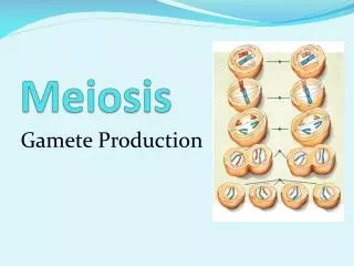
MEIOSIS 4.2
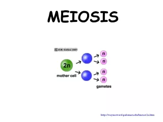
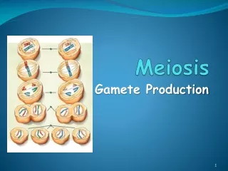
Key Concepts • Gametes have half the number of chromosomes that body cells have. • During meiosis, diploid cells undergo two cell divisions that result in haploid cells.
Haploid and Diploid • The cells which contain two of each type of chromosome are called diploid. • The cells which contain one of each type of chromosome are called haploid. • In diploid cells each pair of chromosomes have the same genes, arranged in the same sequence. However, they do not usually have the same alleles of all of these genes. They are therefore not identical but instead are homologous.
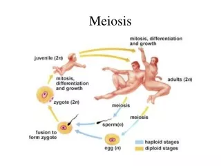
Meiosis • The number of chromosomes in a cell can be reduced from diploid to haploid by the process of meiosis. • Living organisms that reproduce sexually have to halve their chromosome number at some stage in the life cycle because the fusion of gametes during fertilization doubles it.
Homologous Chromosomes • In a diploid human cell, the 46 chromosomes can be grouped into 23 pairs of chromosomes called homologous chromosomes. • Homologous means similar in shape and size and it means that the two chromosomes carry the same genes. The reason there are two of each is that one came from the father and the other from the mother. • Although a pair of chromosomes carry the same genes, they are not identical, because the alleles for the genes from each parent could be different.
4.2.1State that meiosis is a reduction division of a diploid nucleus to form haploid nuclei. • Meiosis is a form of cell division which results in the formation of gametes. • One characteristic feature which makes meiosis unique is that each new cell which results from meiosis has only half the number of chromosomes that a typical cell in that organism has. For example, humans have 46 chromosomes in their cells, but in the gametes (sperm and egg cell) there are only 23 chromosomes. • Cells with half the number of chromosome number are called haploid cells.
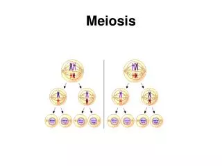
4.2.3 Outline the process of meiosis including pairing of homologous chromosomes and crossing over, followed by two divisions, which results in four haploid cells. • The phases of meiosis • Meiosis is a step-by step process by which a diploid parent cell produces four haploid daughter cells. • In order to produce four cells, the parent cell must divide two times: the first meiotic division makes two cells and then each of these divides during the second meiotic division to make a total of four cells. • http://www.youtube.com/watch?v=D1_-mQS_FZ0 • Video on meiosis • http://www.johnkyrk.com/mitosis.html • animation of meiosis and mitosis • http://www.layyous.com/Videoclips/meiosis%20.htm • meiosis video
Interphase • Interphase • Meiosis is preceded by an interphase, during which the chromosomes duplicate. At the end of this interphase, each chromosome consists of two genetically identical sister chromatids attached together, but at this stage the chromosomes are not visible under the microscope except as a mass of chromatin. The cell’s centrosomes have also duplicated by the end of this stage.
Prophase I • Prophase I is the most complex phase of meiosis. • Early in this phase, the chromatin coils up, so that individual chromosomes become visible with the microscope. • In a process called synapsis, homologous chromosomes, each composed of two sister chromatids, come together as pairs. The resulting structure, consisting of four chromatids , is called a tetrad. • During synapsis, chromatids of homologous chromosomes exchange segments in a process called crossing over. Crossing over rearranges genetic information. Crossing over can make an important contribution to the genetic variability resulting from sexual reproduction.
Prophase I • As prophase I continues, chromosomes condense further as the nucleoli disappear. • Now the centrosome move away from each other, and a spindle starts to form between them. The nuclear envelope breaks into fragments. The chromosome tetrad, captured by spindle microtubule, are moved toward the center of the cell.
Metaphase -I • At metaphase I, the chromosome tetrad are aligned on the metaphase plate, midway between the two poles of the spindle. • Each chromosome is condensed and thick, with its sister chromatid still attached at their centromeres. • Spindle microtubules are attached to kinetochores at the centromeres. • The homologous chromosomes of each tetrad are poised to move toward opposite poles of the cell.
Anaphase I • Anaphase I of meiosis marked by the migration of chromosomes toward the two poles of the cell. • Spindle fibres from the poles attach to chromosomes and pull them to opposite poles of the cell. • The sister chromatids making up each doubled chromosomes remain attached at their centromeres. Only the tetrads split up. ( we can see doubled chromosomes , the haploid number moving toward each spindle pole.
Telophase I • In telophase I, the chromosomes arrive at the poles of the cell. • Each pole of the cell has a haploid chromosome set, though it is duplicate form that is each chromosome consists of two sister chromatids. Usually cytokinesis occurs along with Telophase I, and two haploid daughter cells are formed. • Spindle and spindle fibre disintegrates • Usually, the chromosomes uncoil and new nuclear membrane forms and there is an interphse before meiosis II begins and this happens in some species and in other species daughter cells produced in the first meiotic division immediately begins preparation for the second meiotic division. • In either case no chromosome duplication occurs between Telophase I and the onset of meiosis II • Now meiosis II takes place to separate the sister chromatids.
Prophase II and Metaphase II • Prophase II • DNA condenses into visible chromosomes again. • New meiotic spindle fibres are produced • Metaphase II • Nuclear membrane disintegrate. • The individual chromosomes line up along the equator of each cell in no special order; this is called random orientation. • Spindle fibres from opposite poles attach to each of the sister chromatids at the centromere.
Anaphase II and Telophase II • Anaphase II • Centromeres of each chromosome split, relasing each sister chromatid as an individual chromosome. • The spindle fibre pull individual chromatids to opposite ends of the cell. • In animal cells, cell membranes pinch off in the middle, whereas in plant cells new cell plates form to demarcate the four cells. • Telophase II • Chromosomes unwind their strands of DNA • Nuclear envelopes form around each of the four haploid cells preparing them for cytokinesis.
4.2.4 Explain that non-disjunction can lead to changes in chromosomes number, illustrated by reference to Down syndrome (trisomy 21) • Sometimes chromosomes do not separate the way they are expected to during the first or second meiotic division. This results in an unequal distribution of chromosomes. In humans, it means that an egg cell or sperm cell might have 24 instead of 23 chromosomes. • This unexpected distribution of chromosomes is due to a non-disjunction, a process by which two or more homologous chromosomes stick together instead of separating. • In the case of Down’s syndrome, non-disjunction happens in the 21st pair of chromosomes: the child receives 3 instead of 2. Such an anamoly is called trisomy and Down’s syndrome is also referred to as trisomy 21.
Down’s Syndrome • Having an additional chromosome brings about malformation of the digestive system and causes differing degrees of learning difficulties. • Children with Down’s syndrome follow specialized education programmes adapted for their needs. • The risk of Down’s syndrome increases as the age of the mother increases, particularly over the age of 35. • Non-disjunction can happen with other chromosomes, and all of them can have a major impact on a child’s development. • Some developmental consequences are so severe that the fetus may not survive beyond a few weeks or months.
Analysis of the human karyotype (above To determine if the individual is a male or female, the last pair of chromosomes must be examined. In this case, there is a big x chromosome and a small Y chromosome to indicate that it is a male. • To see if non-disjunction has occurred, the pairs of chromosomes must be checked to see if there are more or fewer than two for any pair. In this case, the 21st pair has three chromosomes indicating Down’s syndrome. • What cannot be determined from this information is how severely the boy will be affected.
Down’s Syndrome (Additional Information) • Most children with Down syndrome have some of the following physical traits: • Short stature. A child often grows slowly and, as an adult, is shorter than average. • Weak muscles (hypotonia) throughout the body. A child may seem to have less strength than other children of the same age. Weak abdominal muscles also make the stomach stick out. Normally, children's stomach muscles gradually strengthen around age 2. • A short, wide neck with excess fat and skin. Usually, this trait is less obvious as the child gets older.
Short, stocky arms and legs. Some children also have a wide space between the big toe and second toe. • A single crease across the center of the palms of the hands. This is called a transverse palmar crease or simian line.
Facial Features • Down syndrome often results in distinct facial features, such as: • Small, low-set ears. • Irregularly shaped mouth and tongue. The child's tongue may partly stick out. The roof of the mouth (palate) may be narrow and high with a downward curve. • A nasal bridge that looks pushed in. The nasal bridge is the flat area between the nose and eyes. • Tissue buildup on the colored part of the eye (iris). These areas are known as Brushfield's spots and do not affect the child's vision. • Irregular and crooked teeth that often come in late and not in the normal sequence.
-- • A child may have other medical conditions related to Down syndrome, such as: • Cognitive disability (mental retardation). Most children with Down syndrome have mild to moderate cognitive disability. • Heart defects. About half of children with Down syndrome are born with a heart defect.1 Most defects are diagnosed at birth or shortly thereafter. • Diseases such as hypothyroidism, celiac disease, and eye conditions. • Children with Down syndrome are also prone to developing other health problems. For example, respiratory infections, hearing problems, and dental problems are common.
4.2.5 State that, in karyotyping, chromosomes are arranged in pairs according to their size and structure. • A karyotype is a photograph of the chromosomes found in a cell arranged according to a standard format. The chromosomes are placed in order according to their size and shape. The shape depends mainly on the position of the centromere. • The number and appearance of the chromosomes in an organism is called the karyotype. Living organisms that are members of the same species usually have the same karyotype.
Karyotype • A karyotype is made by the following steps • The cells are stained and prepared on a glass slide to see their chromosomes under a light microscope. • Photomicrograph images are obtained of the chromosomes during mitotic metaphase. • The images are cut out and separated, a process which can be done using scissors or using a computer. • The images of each pair of chromosomes are placed in order by size and the position of their centromeres.
State that karyotyping is performed using cells collected by chorionic villus sampling or amniocentesis, for pre-natal diagnosis of chromosome abnormalities.- • http://video.about.com/pregnancy/Amniocentesis.htm Amniocentesis
Amniocentesis • From a karyotype, the gender of a person can be deduced and chromosome abnormalities can be detected. The most useful time to do this before birth. Cells have to be obtained from the fetus. There are two ways of doing this. • Amnicentesis • A sample of amniotic fluid is removed from the amniotic sac around the fetus. To do this, a hypodermic needle is inserted through the wall of the mother’s abdomen and wall of the uterus. Amniotic fluid is drawn out into a syringe. It contains cells from the fetus.
Chorionic villus sampling • Cells are removed from the fetal tissues in the placenta called chorionic villi. • As with amniocentesis a hypodermic needle, inserted through the mother’s abdomen and uterus wall, is used to obtain the cells. • Once fetal cells have been obtained, they are incubated with chemicals that stimulate them to divide by mitosis. Another chemical is used which stops mitosis in metaphase of mitosis. Chromosomes are most easily visible in metaphase. A fluid is used to burst the cells and spread out the chromosomes. The burst cells are examined using a microscope and a photograph is taken of the chromosomes from one cell. The chromosomes in the photograph are cut out and arranged into pairs according to their size and structure. This is called karyotyping.
4.2.7Analyse a human karyotype to determine gender and whether non-disjunction has occurred. • http://www.biology.arizona.edu/human_bio/activities/karyotyping/karyotyping.html • (karyotyping activity) • http://www.biology.arizona.edu/human_bio/activities/karyotyping/patient_c/patient_c.html • (karyotyping activity) • The gender of the fetus can be determined form the sex chromosomes.
The preparation of a karyotype is an expensive and invasive procedure. It is usually used for seeing if an unborn baby has any chromosomal anomalies: 45 or 47 chromosomes instead of 46. If the parents or doctors are concerned about the chromosomal integrity of an unborn child (for example, if an expected mother is over the age of 35), a karyotype is recommended.
Karyotypes can be analysed to find out whether a fetus has chromosomal abnormalities.. sometimes chromosomes that should separate and move to opposite poles during meiosis do not separate and instead move to the same pole. This can happen in either the first or the second division of meiosis. Non-separation of chromosomes is called non-disjunction. The result is that gametes are produced with either one chromosome too many or too few.