Download
1 / 1
10 likes | 96 Vues
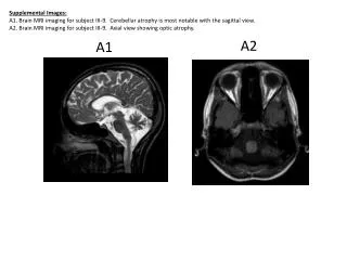
Explore MRI images revealing cerebellar atrophy in the sagittal view and optic atrophy in Subject III-9. Detailed analysis and significance discussed.

E N D
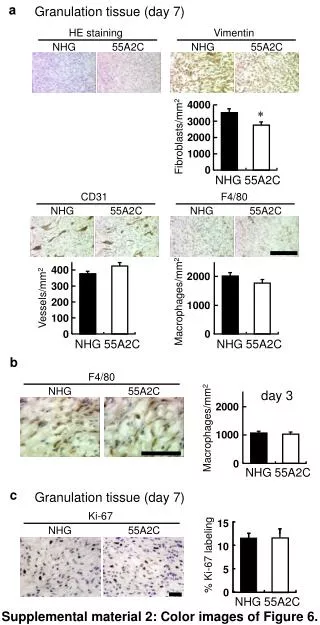
Supplemental Images: A1. Brain MRI imaging for subject III-9. Cerebellar atrophy is most notable with the sagittal view. A2. Brain MRI imaging for subject III-9. Axial view showing optic atrophy. A2 A1