Download
1 / 100
1k likes | 1.44k Vues
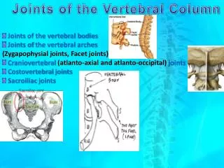
Joints of the Vertebral Column. Joints of the vertebral bodies Joints of the vertebral arches (Zygapophysial joints, Facet joints) Craniovertebral (atlanto-axial and atlanto-occipital) joints Costovertebral joints Sacroiliac joints .

E N D
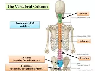
Joints of the Vertebral Column • Joints of the vertebral bodies • Joints of the vertebral arches • (Zygapophysial joints, Facet joints) • Craniovertebral (atlanto-axial and atlanto-occipital) joints • Costovertebral joints • Sacroiliac joints
A typical vertebra has 6 joints with adjacent vertebrae. 4 synovial joints (2 above & 2 below) 2 symphyses (1 above & 1 below) Each symphysis includes an intervertebral disc.
JOINTS OF VERTEBRAL BODIES Symphyses(Secondary cartilaginous joints) Designed for weight-bearing and strength. The articulating surfaces of adjacent vertebrae are connected by intervertebral discs and ligaments. The intervertebral disc consists of an outer anulus fibrosus, which surrounds a central nucleus pulposus.
Anulus fibrosus consists of an outer ring of collagen surrounding a wider zone of fibrocartilage arranged in a lamellar configuration. This arrangement of fibers limits rotation between vertebrae. Nucleus pulposus (L. pulpa, fleshy) core of the intervertebral disc. Fills the center of the intervertebral disc, is gelatinous, and absorbs compression forces between vertebrae. Their semifluid nature is responsible for much of the flexibility and resilience of the intervertebral disc/column.
Intervertebral discs Provide strong attachments between the vertebral bodies Unite them into a continuous semirigid column Form the inferior half of the anterior border of the IV foramen. In aggregate, the discs account for 20-25% of the length (height) of the vertebral column.
No intervertebral disc between C1 & C2 Most inferior functional disc is between L5 & S1 Thickness of the discs increases - Vertebral column descends. Relative thickness (Disc thickness/Body size)- Range of movement Most clear--- Cervical & Lumbar regions Disc thickness most uniform in thoracic region. The discs are thicker anteriorly in the cervical and lumbar regions, their varying shapes producing the secondary curvatures of the vertebral column.
Function of the Intervertebral Discs The semifluid nature of the nucleus pulposus allows it to change shape and permits one vertebra to rock forward or backward on another, as in flexion and extension of the vertebral column.
A sudden increase in the compression load on the vertebral column causes the semifluid nucleus pulposus to become flattened. The outward pushing of the nucleus is accommodated by the resilience of the surrounding anulus fibrosus.
Sometimes, outward push is too great for anulus fibrosus It ruptures Allows nucleus pulposus to herniate Protrude into the vertebral canal, where it may press on the spinal nerve roots, spinal nerve, or even the spinal cord.
With advancing age, the water content of the nucleus pulposus diminishes and is replaced by fibrocartilage. The collagen fibers of the anulus degenerate and, as a result, the anulus cannot always contain the nucleus pulposus under stress. In old age the discs are thin and less elastic, and it is no longer possible to distinguish the nucleus from the anulus. 66 year old male 20 y old male
JOINTS OF VERTEBRAL ARCHES (ZYGAPOPHYSIAL JOINTS-FACET JOINTS Plane synovial joints between superior and inferior articular processes of adjacent vertebrae. Those in the cervical region are especially thin and loose, reflecting the wide range of movement. Accessory ligaments unite the laminae, transverse processes, and spinous processes and help stabilize the joints.
Zygapophysial joints permit gliding movementsbetween the articular processes. • Shape and disposition of the articular surfaces determine the types of movement possible.
«UNCOVERTEBRAL» JOINTS Clefts (of Luschka UNCINATE PROCESS Lateral margins of the upper surfaces of typical cervical vertebrae; elevated into crests or lips.
May articulate with the body of the vertebra above to form small "uncovertebral" synovial joints. Commonly develop between the unci of the bodies of C3 or 4-C6 or 7. @ lateral and posterolateral margins of the intervertebral discs. Considered as synovial joints by some; others; by others as degenerative spaces (clefts) in the discs occupied by extracellular fluid. 1= First rib 2 = Vertebral body of C7 3 = Spinous processes 4 = Uncinate process 5 = Uncovertebral (apophyseal or Luschka's) joint
LIGAMENTS Joints between vertebrae are reinforced and supported by numerous ligaments, which pass between vertebral bodies and interconnect components of the vertebral arches. Anterior and posterior longitudinal ligaments On the anterior and posterior surfaces of the vertebral bodies. Extend along most of the vertebral column.
Anterior longitudinal ligament Attached superiorly to the base of the skull Extends inferiorly to the anterior surface of the sacrum Along its length it’s attached to vertebral bodies & intervertebral discs.
Posterior longitudinal ligament On the posterior surfaces of vertebral bodies Lines the anterior surface of the vertebral canal Attached along its length to vertebral bodies &intervertebral discs. Tectorial membrane Upper part of posterior the longitudinal ligament Connects C2 to intracranial aspect of the base of the skull
Ligamenta flava • Between the laminae of adjacent vertebrae on each side. • Thin, broad ligaments , and of elastic tissue • Form part of the posterior surface of the vertebral canal. • Runs between posterior surface of the lamina on the vertebra below to the anterior surface of the lamina of the vertebra above. • Resist separation of the laminae in flexion • Assist in extension back to anatomical position.
Supraspinous ligament Along the tips of the spinous processes from C7 to the sacrum
Ligamentum nuchae • From C7 to the skull • A triangular, sheet-like structure in the median sagittal plane. • Base of the triangle attached to the skull • Apex attached to the tip of the spinous process of C7 • Deep side of the triangle attached to the posterior tubercle of C1 & spinous processes of the other cervical vertebrae • Supports the head • Resists flexion & facilitates returning head to anatomical position
Interspinous ligaments (Interspinal ligaments) • Between adjacent vertebral spinous processes. • Attach from the base to the apex of each spinous process • Blend with the supraspinous ligament posteriorly • Blend with the ligamenta flava anteriorly • on each side.
CRANIOVERTEBRAL JOINTS 2 sets of joints Atlanto-occipital joints Between atlas(C1) & occipital bone of the cranium Atlanto-axial joints Between atlas and axis (C2) A wider range of movement than in rest of the vertebral column. Articulations: Occipital condyles, Atlas& Axis. • Synovial joints without intervertebral discs
Atlanto-occipital Joints Articulations between Superior articular surfaces of the lateral masses of the atlas & Occipital condyles Synovial joints of the condyloid type
Atlanto-occipital Joints Nodding of the head, flexion and extension of the head occurring when indicating approval (the “yes” movement). Sideways tilting of the head. Main movement Flexion, with a little lateral flexion & rotation
The cranium and C1 are also connected by • Anterior & posterior atlanto-occipital membranes • Extend from the anterior and posterior arches of C1 to the anterior and posterior margins of the foramen magnum. • Help prevent excessive movement of atlanto-occipital joints.
Atlanto-axial Joints 3 atlanto-axial articulations 2 (right & left) lateral atlantoaxial joints – plane type joint between inferior facets of lateral masses of C1 & superior facets of C2 1 median atlantoaxial joint – pivo type joint between dens of C2 & anterior arch of atlas
Atlanto-axial Joints Head turns from side to side, disapproval (“no” movement). Cranium and C1 rotate on C2 as a unit. .
Atlanto-axial Joints During rotation of the head, dens of C2 axis or pivot held in a socket or collar formed Anteriorly by anterior arch of the atlas Posteriorly by transverse ligament of the atlas A strong band extending between tubercles on the medial aspects of lateral masses of C1
Ligaments Superior and inferior longitudinal bands Apical ligament Alar ligaments Cruciate ligament of the atlas Tectorial membrane (Membrana tectoria)
Movements of the Vertebral Column Range of movement Region & individual The mobility primarily from the intervertebral discs. The normal range of movement possible in healthy young adults is typically reduced by 50% or more as they age. Although the movement between any two vertebrae is limited, the summation of movement among all vertebrae results in a large range of movement by the vertebral column.
Movements by the vertebral column • Flexion • Extension • Lateral flexion • Rotation • Circumduction • The range of movement of the vertebral column is limited by the: • Thickness, elasticity, and compressibility of the IV discs • Shape and orientation of the zygapophysial joints • Tension of the joint capsules of the zygapophysial joints • Resistance of the back muscles and ligaments (e.g., the ligamentaflava and the posterior longitudinal ligament) • Attachment to the thoracic (rib) cage • Bulk of surrounding tissue.
CLINICAL NOTES
Disc Hernia & Back Pain A tear within the anulus fibrosus Material of the nucleus pulposus can track This material tracks into the vertebral canal or into the intervertebral foramen Pressure on neural structures. This is a common cause of back pain.
Discectomy/laminectomy A prolapsed intervertebral disc may impinge upon the meningeal (thecal) sac, cord, and most commonly the nerve root, producing symptoms attributable to that level. Neurological signs- Surgery It is of the utmost importance that the level of the disc protrusion is identified before surgery. This may require MRI scanning and on-table fluoroscopy.
PELVIS L. Basin Part of the trunk inferoposterior to the abdomen Area of transition between the trunk and the lower limbs Pelvic cavity Inferiormost part of the abdominopelvic cavity. Anatomically, the pelvis is the part of the body surrounded by the pelvic girdle (bony pelvis), part of the appendicular skeleton of the lower limb.
Pelvis is subdivided into greater and lesser pelves. Greater pelvis Surrounded by the superior pelvic girdle. Occupied by inferior abdominal viscera, affording them protection. Lesser pelvis Surrounded by the inferior pelvic girdle, which provides the skeletal framework for both the pelvic cavity and the perineum—compartments of the trunk separated by the musculofascial pelvic diaphragm.
A basin-shaped ring of bones that connects the vertebral column to the two femurs. • Primary functions of the pelvic girdle: • Bear the weight of the upper body when sitting and standing. • Transfer that weight from the axial to the lower appendicular skeleton for standing and walking. • Provide attachment for the powerful muscles of locomotion and posture and those of the abdominal wall. PELVIC GIRDLE
The pelvic bone is irregular in shape and has two major parts separated by an oblique line on the medial surface of the bone: • pelvic bone above this line represents lateral wall of the false pelvis, part of the abdominal cavity. • pelvic bone below this line represents the lateral wall of the true pelvis, contains the pelvic cavity. • Linea terminalis lower two-thirds of this line & contributes to the margin of the pelvic inlet.
Pelvic girdle is formed by 3 bones: • Right and left hip bones (coxal bones; pelvic bones): large, irregularly shaped bones, each of which develops from the fusion of three bones • Ilium • Ischium • Pubis • Sacrum: formed by the fusion of five, originally separate, sacral vertebrae.
In infants and children, hip bones are 3 separate bones united by a triradiate cartilage at the acetabulum, the cup-like depression in the lateral surface of the hip bone, which articulates with the head of the femur. After puberty, the ilium, ischium, and pubis fuse to form the hip bone. The two hip bones are joined anteriorly at the pubic symphysis (L. symphysis pubis) and articulate posteriorly with the sacrum at the sacroiliac joints to form the pelvic girdle.
Ilium Superior, fan-shaped part of the hip bone Ala, or wing, of the iliumspread of the fan Body of the ilium, the handle of the fan. On its external aspect, the body participates in formation of the acetabulum.
Ilium The entire superior margin of the ilium is thickened to form a prominent crest (iliac crest) terminates anteriorly as the anterior superior iliac spine and posteriorly as the posterior superior iliac spine.
A prominent tubercle, tuberculum of iliac crest, projects laterally near the anterior end of the crest; the posterior end of the crest thickens to form the iliac tuberosity. Inferior to the anterior superior iliac spine, rounded protuberance called anterior inferior iliac spine.
Ilium Posteriorly, the sacropelvic surface of the ilium has an auricular surface and an iliac tuberosity articulation with sacrum.
Ischium Has a body and ramus (L. branch). Body of the ischium forms the acetabulum Ramus of the ischium forms part of the obturator foramen.
Ischium Ischial tuberosity: large posteroinferior protuberance of ischium Ischial spine: Small pointed posteromedial projection near the junction of the ramus and body Lesser sciatic notch: Concavity between the ischial spine and the ischial tuberosity Greater sciatic notch: Larger concavity superior to the ischial spine and formed in part by the ilium.