Download
1 / 14
160 likes | 438 Vues
Human Skeleton. Human Skeleton. Human Skeleton. Skeletal System #1. Functions- Support & movement, muscle attachment, levers Protection of internal organs Manufacture of blood cells Storage of minerals, phosphorous, calcium. Bone Structure- #2 Figure 1- Typical Long Bone. 1. Cartilage

E N D
Skeletal System #1 • Functions- • Support & movement, muscle attachment, levers • Protection of internal organs • Manufacture of blood cells • Storage of minerals, phosphorous, calcium
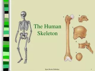
Bone Structure- #2Figure 1- Typical Long Bone • 1. Cartilage • Located at the ends of bones, reduces friction • 2. Spongy bone • Located inside compact bone & at the ends of long bones, many air spaces • 3. Marrow • Soft tissue located in the center of hollow bones, produce red blood cells & some white blood cells
Bone Structure- #2Figure 1- Typical Long Bone • 4. Periosteum • Tough membrane that encases the bone • 5. Compact bone • Dense bone, strong, located along the center of long bones • 6. Ligament or Tendon • Ligament- connects bone to other bone • Tendon- connects muscle to bone
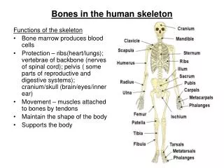
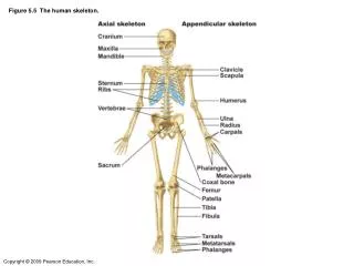
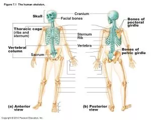
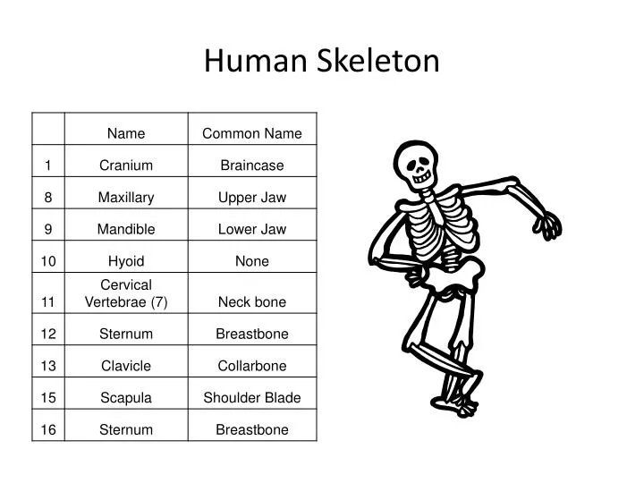
Bone Structure- #3Figure 2 • a. Vertebral column • Protects spinal cord • b. Thoracic cavity or Ribcage • Protects lungs & heart • c. Cranium • Protects brain • d. Mandible • Shapes face, allows chewing
Bone Structure- #3Figure 2 • e. Shoulder girdle • Supports, anchors arm • f. Pelvis • Protects pelvic organs, anchors leg • g. Cartilage • Reduces friction between bones • h. Spongy bone • Bone with air spaces
Bone Structure- #3Figure 2 • i. Marrow • Forms blood cells • j. Compact bone • Gives bone strength and hardness • k. Blood vessels/nerves • l. Osteocyte • Living bone cells • m. Haversian Canal • Canal for blood vessels in bone
Bone Development- #4 • Ossification • The changing of cartilage to bone • Osteocytes deposit minerals that replace cartilage forming bone
Cells Associated Tissue- #5 • Osteocytes • living bone cells, deposit or absorbs minerals, bone growth and ossification • Cartilage- 2 types • Temporary cartilage- ossifies into bone • Permanent cartilage- remain cartilage through out your life • ends of bones, nose, ear
Cells Associated Tissue- #5 • Tendons • Hold muscle to bone • Ligaments • Hold bone to bone
Joints- #6 • Point where bones meet • Types- • Fixed- immovable, no movement, cranium • Semimovable- allows some motion, vertebrae • Freely movable- allows much motion
Joints- #6 • Movable- allows much motion • Ball & socket- shoulder, hip • Hinge- knee, elbow • Pivot- top 2 vertebrae, elbow • Gliding- wrist, foot • Saddle- base of thumb