Download
1 / 23
331 likes | 636 Vues
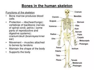
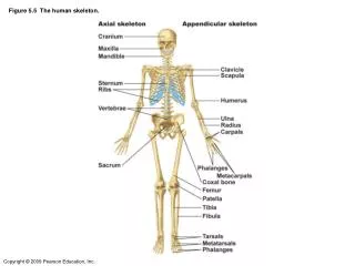
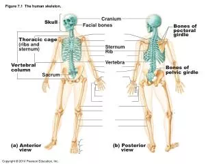
The Human Skeleton. Axial Skeleton. Axial Skeleton. Skull. Sternum. Ribs. VertebralColumn. Skull. Divided into two parts: a) Calvaria b) Face . a) Calvaria. Parietal Bone. Frontal Bone. Occipital Bone. Temporal Bone. Calvaria Cont.

E N D
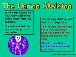
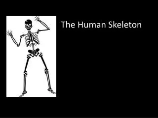
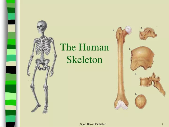
The Human Skeleton Sport Books Publisher
Axial Skeleton Sport Books Publisher
Axial Skeleton Skull Sternum Ribs VertebralColumn Sport Books Publisher
Skull • Divided into two parts: a) Calvaria b) Face Sport Books Publisher
a) Calvaria Parietal Bone Frontal Bone Occipital Bone Temporal Bone Sport Books Publisher
Calvaria Cont. • May be fractured in blows to the skull (e.g., in hockey, being checked and hitting the skull on the ice) • Temporal bone: • more fragile of the calvaria bones • overlies one of the major blood vessels • if fractured and displaced internally = medical emergency (picture) Sport Books Publisher
b) Facial Bones Lacrimal Bone Nasal Bone Zygomatic Bone Maxilla Bone Mandible Bone Sport Books Publisher
Facial Bones Cont’d • Often broken in contact sports due to rough impact • Some fractures across the maxilla (upper jaw) can leave the lower face separated from the upper face Sport Books Publisher
Lumbar vertebra, lateral view 7 Cervical Vertebrae (of the neck) 12 Thoracic Vertebrae (of the chest) Lumbar vertebra, superior view 5 Lumbar Vertebrae (of the lower back) Sacrum (mid-line region of buttocks) Coccyx (4 or 5 fused vertebrae of the tail bone) Vertebral Column Sport Books Publisher
Vertebral Column • Vertebrae are arranged in a cylindrical column interspersed with fibrocartilaginous (intervertebral) discs • Function: • provides a strong and flexible support for the body and the ability to keep the body erect • the point of attachment for the muscles of the back. • protect the spinal cord and nerves • absorbs shock through the intervertebral discs without causing damage to other vertebrae Sport Books Publisher
Ribs • Twelve pairs • Made up of : • bone • cartilage which strengthen the chest cage and permit it to expand. • Curved and slightly twisted making it ideal to protect the chest area Sport Books Publisher
Ribs Cont’d • All 12 pairs of ribs articulate with the twelve thoracic vertebrae posteriorly • Classified into three groups based on anterior attachment: (picture) • true ribs • 1-7 • attach to both the vertebrae and the sternum • false ribs • 8-10 • attach only to the sternum indirectly, through 7th rib • floating ribs • 11 and 12 • only attach to the vertebral column Sport Books Publisher
True Ribs (1-7) False Ribs (8-10) Floating Ribs (11-12) The Ribs Manubrium Sternal Body Xiphoid Process Costal Cartilages Sport Books Publisher
Sternum • Mid-line breast bone • The clavicles and ribs one to seven articulate with the sternum Sternum – comprised of the manubrium, sternal body and xiphoid process Sport Books Publisher
Appendicular Skeleton Sport Books Publisher
Appendicular skeleton Consists of: • 1. The pectoral gridle (chest) • 2. Pelvic girdle (hip) • 3. The upper limbs • 4. The lower limbs Sport Books Publisher
Clavicle Scapula 1.Pectoral Girdle Consists of: • Scapula (shoulder blade) • Clavicle (collar bone) • Allows the upper limb great mobility • The sternoclavicular joint is the only point of attachment between the axial skeleton and the pectoral girdle Sport Books Publisher
2. Pelvic Girdle • Formed by pair of os coxae (hip bones) • supports the bladder and abdominal contents • Attachment: • Posteriorly – join with the sacrum • Anteriorly - join to each other anteriorly • Laterally – join to the head of thigh bone through a cup-shaped acetabulum Sport Books Publisher
Humerus Radius Ulna 3. Upper Limb • Humerus • The arm bone • shoulder to elbow • Radius and Ulna • The forearm bones • elbow to wrist • the radius being located on the thumb side of the hand • when you pronate the forearm, the radius is actually crossing over the ulna - try it yourself Sport Books Publisher
Carpals Proximal Phalanx Metacarpals Phalanges Middle Phalanx Distal Phalanx Upper Limb Cont. Sport Books Publisher
Femur Patella 4. Lower Limb • Femur • thigh bone • from hip to knee • Patella • knee cap • sesamoid bone in the tendon of the quadriceps muscles (thigh) Sport Books Publisher
Fibula Tibia Lat. malleolus Med. malleolus Lower Limb Cont’d • Tibia and Fibula • leg bones • From knee to ankle • Tibia is medial and fibula is lateral • Medial malleolus and Lateral malleolus • The distal ends of the tibia and fibula, respectively • commonly referred to as the "ankle bones" • can be easily palpated Sport Books Publisher
Talus Calcaneus Tarsals Metatarsals Phalanges Lower Limb Cont’d • Tarsals • ankle bones • calcaneus or the heel bone • talus • Metatarsals • 5 bones of the foot • unite with the toes • Phalanges • toe bones • three per toe except the big toe - proximal, middle and distal Sport Books Publisher