Download
1 / 26
420 likes | 1.39k Vues
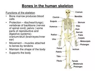
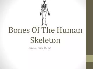
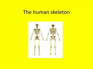
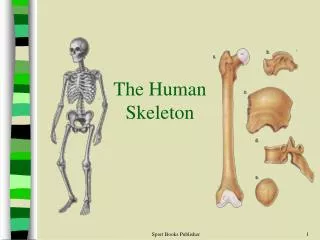
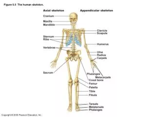
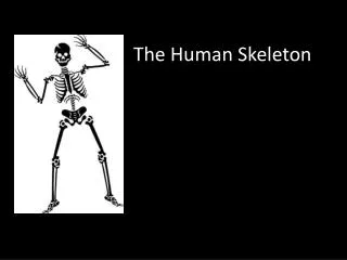
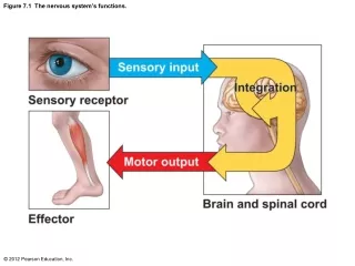
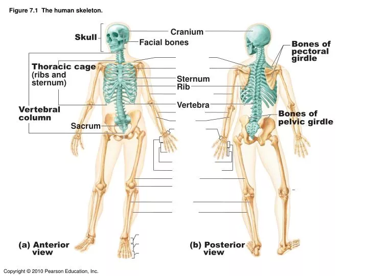
Figure 7.1 The human skeleton. Cranium. Skull. Facial bones. Bones of pectoral girdle. Thoracic cage (ribs and sternum). Sternum. Rib. Vertebra. Vertebral column. Bones of pelvic girdle. Sacrum. (a) Anterior view. (b) Posterior view.

E N D
Figure 7.1 The human skeleton. Cranium Skull Facial bones Bones of pectoral girdle Thoracic cage (ribs and sternum) Sternum Rib Vertebra Vertebral column Bones of pelvic girdle Sacrum (a) Anterior view (b) Posterior view
Figure 7.2a The skull: Cranial and facial divisions and fossae. Bones of cranium (cranial vault) Coronal suture Squamous suture Facial bones Lambdoid suture (a) Cranial and facial divisions of the skull
Figure 7.5a Bones of the lateral aspect of the skull, external and internal views. Sphenoid bone (greater wing) Zygomatic process Alveolar margins Mastoid process Styloid process Mandibular condyle Mental foramen Mandibular ramus Coronoid process (a) External anatomy of the right side of the skull
Figure 7.4b Anatomy of the anterior and posterior aspects of the skull. Sagittal suture Sutural bone External occipital protuberance External occipital crest Occipital condyle (b) Posterior view
Figure 7.6a Inferior aspect of the skull, mandible removed. Maxilla (palatine process) Hard palate Palatine bone Infraorbital foramen Zygomatic bone Sphenoid bone (greater wing) Temporal bone (zygomatic process) Mandibular fossa Temporal bone Occipital condyle (a) Inferior view of the skull (mandible removed)
Figure 7.8 The temporal bone. Squamous region External acoustic meatus Zygomatic process Mandibular fossa Mastoid process Styloid process
Figure 7.9 The sphenoid bone. Lesser wing Greater wing sella turcica Body of sphenoid (a) Superior view Body of sphenoid Lesser wing Greater wing Pterygoid process (b) Posterior view
Figure 7.10 The ethmoid bone. ANTERIOR VIEW Crista galli Olfactory foramina Cribriform plate Ethmoidal sinuses Perpendicular plate Middle nasal concha
Figure 7.15 Paranasal sinuses. Frontal sinus Frontal sinus Ethmoidal sinus Ethmoidal sinus Sphenoid sinus Sphenoid sinus Maxillary sinus Maxillary sinus (b) Medial aspect (a) Anterior aspect
Figure 7.13 Bones that form the orbits. (YOU DON’T NEED TO KNOW ALL THE DETAILS )
Figure 7.18 Structure of a typical vertebra. Posterior Lamina Spinous process Transverse process facet Vertebral foramen Pedicle Body Anterior
Figure 7.20 Posterolateral views of articulated vertebrae. Dens of axis Transverse process C1 (atlas) Transverse costal facet (for tubercle of rib) C2 (axis) C3 Intervertebral disc Body Inferior costal facet (for head of rib) Transverse processes Spinous process C7 (b) Thoracic vertebrae (a) Cervical vertebrae Body Transverse process Intervertebral disc Spinous process (c) Lumbar vertebrae
Figure 7.17a Ligaments and fibrocartilage discs uniting the vertebrae. Intervertebral disc Transverse process Sectioned spinous process Intervertebral foramen Interspinous ligament Sectioned body of vertebra Median section of three vertebrae, illustrating the composition of the discs and the ligaments
Table 7.2 Regional Characteristics of Cervical, Thoracic, and Lumbar Vertebrae (2 of 3)(DON’T NEED TO KNOW ALL DETAILS)
Table 7.2 Regional Characteristics of Cervical, Thoracic, and Lumbar Vertebrae (3 of 3) (DON’T NEED TO KNOW ALL DETAILS)
Figure 7.19 The first and second cervical vertebrae. Posterior Posterior C1 facet Transverse process Transverse foramen facet Transverse foramen Facet for dens (a) Superior view of atlas (C1) (b) Inferior view of atlas (C1) Posterior C2 Spinous process Lamina Pedicle facet Transverse process Dens Body (c) Superior view of axis (C2)
Figure 7.23b Ribs. tubercle of rib Spinous process Shaft Transverse Facet (for Tubercle of rib) Ligaments Neck of rib Body of thoracic vertebra Head of rib Facet (for head of rib) (b) Superior view of the articulation between arib and a thoracic vertebra
Figure 7.21a The sacrum and coccyx. Body of first sacral vertebra Anterior sacral foramina Coccyx (a) Anterior view
Figure 7.22a The thoracic cage. Manubrium Body Sternum True ribs (1–7) Xiphoid process False ribs (8–12) Intercostal spaces Costal cartilage L1 Vertebra Floating ribs (11, 12) (a) Skeleton of the thoracic cage, anterior view
Figure 7.23c Ribs. Facets for articulation with vertebrae tubercle Head Neck Junction with costal cartilage (c) A typical rib (rib 6, right), posterior view
Figure 7.35 Skull of a newborn. (DON’T NEED TO KNOW NAMES OF FONTANELLES) Frontal suture Frontal bone Anterior fontanelle Parietal bone Posterior fontanelle Occipital bone (a) Superior view Parietal bone Frontal bone Sphenoidal fontanelle Posterior fontanelle Temporal bone (squamous portion) Mastoid fontanelle Occipital bone (b) Lateral view