Download
1 / 42
510 likes | 1.76k Vues
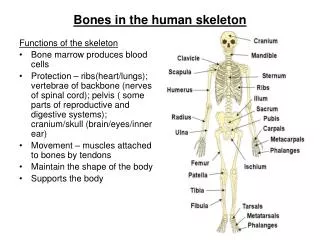
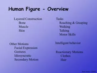
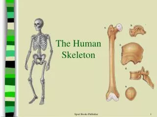
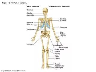
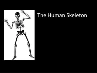
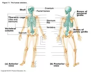
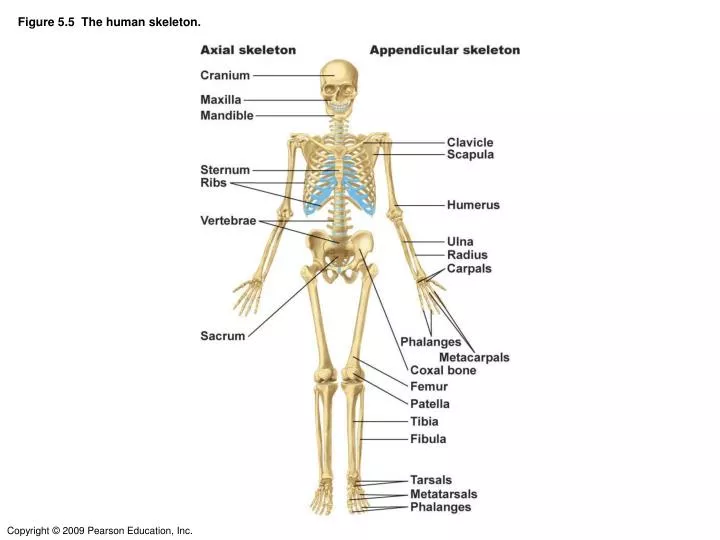
Figure 5.5 The human skeleton. Figure 5.5 The human skeleton. Figure 5.10 Bones of the right side of the pectoral girdle and the right arm and hand. Figure 5.11 Bones of the pelvic girdle and the left leg and foot. Figure 7.24 The pectoral girdle and clavicle. Acromio- clavicular

E N D
Figure 5.10 Bones of the right side of the pectoral girdle and the right arm and hand.
Figure 5.11 Bones of the pelvic girdle and the left leg and foot.
Figure 7.24 The pectoral girdle and clavicle. Acromio- clavicular joint Sternal (medial) end Posterior Clavicle Anterior Acromial (lateral) end (b) Right clavicle, superior view Acromial end Anterior Sternal end Posterior Scapula (a) Articulated pectoral girdle (c) Right clavicle, inferior view
Figure 7.25 The scapula. Coracoid process Acromion Coracoid process Acromion Glenoid cavity Glenoid cavity at lateral angle Spine Medial border (a) Right scapula, anterior aspect (b) Right scapula, posterior aspect Inferior angle Acromion Coracoid process Glenoid cavity Spine (c) Right scapula, lateral aspect
Figure 7.26 The humerus of the right arm Greater tubercle Head of humerus Greater tubercle Lesser tubercle Anatomical neck Surgical neck Deltoid tuberosity Deltoid tuberosity Coronoid fossa Olecranon fossa Medial epicondyle Lateral epicondyle Capitulum Trochlea (a) Anterior view (b) Posterior view
Figure 7.27 Radius and ulna of the right forearm. Radial notch of the ulna Olecranon process Trochlear notch Head Head of radius Coronoid process Neck Radial tuberosity Neck of radius Ulna Radius Radius Styloid process of ulna Styloid process of radius Styloid process of radius (a) Anterior view (b) Posterior view
Figure 7.27d Radius and ulna of the right forearm. Ulnar notch of radius Styloid process Styloid process View Head of ulna (d) Distal ends of the radius and ulna at the wrist
Figure 7.27c Radius and ulna of the right forearm. Olecranon process View Trochlear notch Coronoid process Radial notch (c) Proximal portion of ulna, lateral view
Figure 7.26c Detailed views of articulation at the elbow. Humerus Coronoid fossa Medial epicondyle Capitulum Trochlea Head of radius Coronoid process of ulna Radial tuberosity Radial notch Radius Ulna (c) Anterior view at the elbow region
Figure 7.26d The humerus of the right arm and detailed views of articulation at the elbow. Humerus Olecranon fossa Olecranon process Lateral epicondyle Medial epicondyle Head Neck Ulna Radius (d) Posterior view of extended elbow
Figure 7.28 Bones of the left hand. Phalanges • Distal • Middle • Proximal Metacarpals Carpals Carpals Carpals Radius Ulna Radius (a) Anterior view of left hand (b) Posterior view of left hand
Figure 7.29 Articulated pelvis showing the two hip (coxal) bones (which together form the pelvic girdle), the sacrum, and the coccyx. Sacroiliac joint Iliac crest Anterior superior iliac spine Coxal bone (os coxae or hip bone) Anterior inferior iliac spine llium Sacrum Pubic bone Coccyx Acetabulum Ischium Pubic symphysis Pubic arch
From Wikipedia: The hip bone, innominate bone or coxal bone is a large, flattened, irregularly shaped bone. It has one of the few ball and socket synovial joints in the body – the so called hip joint. It meets its fellow on the opposite side in the middle line in front, and together they form the sides and anterior wall of the pelvic cavity. Together with the sacrum and coccyx, it comprises the pelvis
OS INNOMINATUM The " os innominatum," so named by Galen, is made up of three bones, distinct in childhood, but united in the adult, and termed the "ilium," "ischium," and "pubes." Thus its constituents have received appropriate names, but the bone, consolidated, remains " nameless." Human osteology By Luther Holden 1899
Figure 7.30a Bones of the bony pelvis. Ilium Iliac crest Posterior superior iIiac spine Anterior superior iliac spine Posterior inferior iliac spine Anterior inferior iliac spine Acetabulum Ischial spine Pubis Ischium Inferior ramus of pubis Ischial tuberosity Obturator foramen Ischial ramus (a) Lateral view, right hip bone
Figure 7.30b Bones of the bony pelvis. Body of the ilium (b) Medial view, right hip bone
Figure 7.31 Bones of the right knee and thigh. Neck Greater trochanter Head Lesser trochanter Linea aspera Anterior Lateral condyle Medial condyle Posterior (a) Patella (kneecap) Anterior view Posterior view (b) Femur (thigh bone)
Figure 7.32a The tibia and fibula of the right leg. Lateral condyle Head Medial condyle Tibial tuberosity Anterior crest Fibula Tibia Lateral malleolus Medial malleolus (a) Anterior view
Figure 7.32e The tibia and fibula of the right leg. Parts of fractured fibula (e) X ray of Pott’s fracture of the fibula
Figure 7.33 Bones of the right foot. Talus First metatarsal Phalanges Distal Middle Proximal Calcaneus 1 2 3 4 5 Metatarsals (b) Medial view Tarsals Talus Talus Calcaneus (a) Superior view Calcaneus Fifth metatarsal (c) Lateral view
Joint = Articulation And Body Movement
Table 8.1 Summary of Joint Classes(DON’T NEED TO KNOW ALL DETAILS, BUT KNOW THREE BASIC TYPES OF JOINTS)
(c) Gomphosis (a) Suture (b) Syndesmosis Joint held together with very short, interconnecting fibers, and bone edges interlock. Found only in the skull. Joint held together by a ligament. Fibrous tissue can vary in length, but is longer than in sutures. “Peg in socket” fibrous joint. Periodontal ligament holds tooth in socket. Socket of alveolar process Suture line Fibula Tibia Root of tooth Dense fibrous connective tissue Ligament Periodontal ligament Figure 8.1 Fibrous joints. (DON’T NEED TO KNOW ALL DETAILS)
(a) Synchondroses Bones united by hyaline cartilage Sternum (manubrium) Epiphyseal plate (temporary hyaline cartilage joint) Joint between first rib and sternum (immovable) (b) Symphyses Bones united by fibrocartilage Body of vertebra Fibrocartilaginous intervertebral disc Hyaline cartilage Pubic symphysis Figure 8.2 Cartilaginous joints. (DON’T NEED TO KNOW ALL DETAILS)
Ligament Joint cavity (contains synovial fluid) Articular (hyaline) cartilage Fibrous capsule Articular capsule Synovial membrane Periosteum Figure 8.3 General structure of a synovial joint.
f Nonaxial Uniaxial Biaxial Multiaxial c b Hinge joint (elbow joint) b a e d Figure 8.7b Types of synovial joints.
f Nonaxial Uniaxial Biaxial Multiaxial c b c Pivot joint (proximal radioulnar joint) a e d Figure 8.7c Types of synovial joints.
f Nonaxial Uniaxial Biaxial Multiaxial c b d Condyloid joint (metacarpophalangeal joint) a e d Figure 8.7d Types of synovial joints.
f Nonaxial Uniaxial Biaxial Multiaxial c b e Saddle joint (carpometacarpal joint of thumb) a e d Figure 8.7e Types of synovial joints.
f Nonaxial Uniaxial Biaxial Multiaxial c b f Ball-and-socket joint (shoulder joint) a e d Figure 8.7f Types of synovial joints.
Medial femoral condyle Medial meniscus on medial tibial condyle Patella (f) Photograph of an opened knee joint; view similar to (e) Figure 8.8f The knee joint.
ligament Subacromial bursa Cavity in bursa containing synovial fluid Humerus resting Bursa rolls and lessens friction. Humerus head rolls medially as arm abducts. Humerus moving (b)Showing how a bursa eliminates friction where a ligament (or other structure) would rub against a bone Figure 8.4 Bursae Synovial membrane
Acromion Coracoid process ligament Subacromial bursa ligament Subscapular bursa Greater tubercle of humerus Tendon ligament Scapula Tendon sheath Tendon of long head of biceps brachii muscle (c) Anterior view of right shoulder joint capsule Figure 8.10c The shoulder joint: bursae and tendon sheath
A Closer Look 8.1a Joints: From Knights in Shining Armor to Bionic Humans
A Closer Look 8.1b: Joints: From Knights in Shining Armor to Bionic Humans
Flexion Extension Flexion Extension (d) Angular movements: flexion and extension at theshoulder and knee Figure 8.5d Movements allowed by synovial joints.
Abduction Circumduction Adduction (e) Angular movements: abduction, adduction, andcircumduction of the upper limb at the shoulder Figure 8.5e Movements allowed by synovial joints.
Rotation Lateral rotation Medial rotation (f) Rotation of the head, neck, and lower limb Figure 8.5f Movements allowed by synovial joints.
Pronation (radius rotates over ulna) Supination (radius and ulna are parallel) (a) Pronation (P) and supination (S) Figure 8.6a Special body movements.