0 likes | 79 Vues
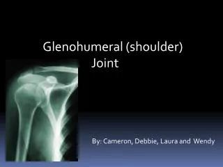
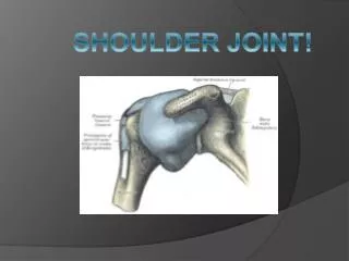
A comprehensive and advanced Shoulder Joint Ultrasound Course designed to provide in-depth knowledge of shoulder anatomy, systematic scanning techniques, pathology detection, and extensive hands-on practice sessions. Participants will learn to accurately assess conditions such as rotator cuff tears, bursitis, tendinopathy, impingement syndromes, and other shoulder pathologies using real-patient case studies. This course is ideal for radiologists, sonographers, physiotherapists, orthopedic specialists, and sports medicine professionals seeking to enhance their musculoskeletal ultrasound diagnos
E N D
Introduction to MSK Ultrasound of the Shoulder Joint Musculoskeletal (MSK) ultrasound visualizes soft tissues in the shoulder joint. It offers real-time imaging, is cost-effective, and radiation-free. This course covers anatomy, scanning methods, and common pathologies. The global MSK ultrasound market is projected to reach $11.3 billion by 2028, reflecting its growing clinical importance. FG
Shoulder Joint Anatomy: Key Structures Bony Landmarks Humerus, scapula, clavicle form the shoulder frame. Rotator Cuff Muscles Supraspinatus Infraspinatus Teres Minor Subscapularis Tendons & Bursa Tendons connect muscles; bursae reduce friction. Sonographic Appearance Normal tissues appear hyperechoic with clear fibrillar patterns.
Ultrasound Transducers and Knobology Transducers 7-15 MHz linear probes provide high-resolution images. Key Settings Adjust depth, gain, focus, and Doppler for optimal imaging. Image Optimization Fine-tuning improves visualization of superficial MSK structures. Proven Technology Linear arrays outperform others for detailed soft tissue exams.
Scanning Techniques: Anterior Approach Patient Positioning Seated with arm in slight external rotation. Transducer Placement Place probe over bicipital groove to visualize biceps tendon. Identifying Structures Locate biceps and subscapularis tendons and glenohumeral joint. Dynamic Assessment Perform movements to check for impingement and tendon movement.
Scanning Techniques: Posterior and Lateral Approaches Posterior Approach Lateral Approach Fluid & Mass Detection Example Evaluate supraspinatus and infraspinatus tendons for tears. Assess acromioclavicular joint and surrounding bursae. Craniocaudal view effectively detects supraspinatus tendon tears. Identify cysts, fluid collections, or masses near tendons.
Common Shoulder Pathologies: Rotator Cuff Tears Partial Thickness Tears Tendon fibers partially disrupted, causing thinning and fluid. Full Thickness Tears Complete tendon discontinuity with retraction visible sonographically. Sonographic Signs Look for hypoechoic gaps, tendon retraction, and surrounding fluid. Accuracy Ultrasound detects tears with 85-95% accuracy.
Common Shoulder Pathologies: Other Conditions Subacromial-subdeltoid Bursitis Biceps Tendinopathy Tendon thickening and irregularity due to overuse or injury. Inflammation causing fluid in bursa, seen as anechoic area. 1 2 4 3 Calcific Tendinitis Joint Effusion Calcium deposits appearing as hyperechoic foci with shadowing. Fluid accumulation in the glenohumeral joint space.
Conclusion: Integrating MSK Ultrasound in Practice POCUS Benefits Quick bedside diagnosis improves treatment efficiency and outcomes. Diagnostic Accuracy Ultrasound guidance increases injection accuracy by 30-40%. Further Learning Advanced courses and resources enhance skills and knowledge. Impact Integrating ultrasound elevates patient care and clinical confidence. visit us ;- https://www.mediflick.com/courses/MSK-Ultrasound-Shoulder-Joint- 67e3b83bd1e1f85a0f813413