Download
1 / 18
210 likes | 1.17k Vues
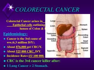
THE REGISTRATION OF COLORECTAL CANCER. The anatomy of the colon rectum and anus,the pathology and treatment of colorectal cancer, and the collection of data on colorectal cancer. FIVE FACTS ABOUT COLORECTAL CANCER. Incidence:

E N D
THE REGISTRATION OF COLORECTAL CANCER The anatomy of the colon rectum and anus,the pathology and treatment of colorectal cancer, and the collection of data on colorectal cancer.
FIVE FACTS ABOUT COLORECTAL CANCER • Incidence: 1 in 14 men and 1 in 20 women in the TCR area develop colorectal cancer during their lifetime. Incidence rates increase with age. • Survival: For people in the TCR area the 5 yr survival estimates are: 42% for colon tumours, 45% for rectal tumours. For anal tumours the figures are 47% for men and 60% for women. • Most common agegroup: 75-80 yrs • Population most at risk: Colon and rectum cancers are most common in developed countries. Anal cancer is most common in patients with HIV and sexually transmitted disease, especially in homosexual men. • Predisposing factors: Western diet, hereditary factors (e.g. familial polyposis coli), inlammatory diseases (e.g. Chrohn’s)
The large intestine is a tube of smooth muscle about 140 cm (4ft 6ins) long joining the ileum (at the ileocaecal valve) to the external surface of the body (at the anus). It is lined with mucous membrane which: Absorbs water and salts from the liquid contents of the ileum, to form faeces. Secretes mucous to facilitate the passage of faeces. Contains neuroendocrine cells to control the function of the intestine The muscular tube then expels the faeces at the anus. THE ANATOMY & FUNCTION OF THE COLON, RECTUM & ANUS
DIAGRAM TO SHOW THE VARIOUS LAYERS OF THE WALL OF THE LARGE INTESTINE
THE LARGE INTESTINE The hepatic flexure The splenic flexure Transverse colon Descending colon Rectosigmoid junction Sigmoid colon Rectum Anus The ascending colon The caecum The appendix
The large intestine is closely packed into the abdominal and pelvic cavities, along with the loops of the small intestine, and urogenital organs. It lies below the stomach. THE ANATOMY OF THE LARGE INTESTINE Stomach Large bowel Small bowel
The parts of the colon: Caecum Ascending colon Hepatic flexure Transverse colon Splenic flexure Descending colon Sigmoid colon Rectosigmoid junction Rectum Anus Cancer registries treat each individual part of the large intestine as a separate tumour site. This means that a patient with tumours in: - the caecum and - ascending colon will be registered for 2 malignancies irrespective of tumour type. A patient with two separate tumours in the ascending colon will be registered for: - a single malignancy if the tumours both have the same morphology, and - 2 separate malignancies if the morphologies are different. THE REGISTRATION OF MALIGNANCIES OCCURING IN THE LARGE INTESTINE N.B. Many clinicians regard the colon and rectum as a single organ - the large intestine.This may lead to duplicate cancer registrations when a rectal tumour is loosely referred to as “colon cancer”.
Most colorectal malignancies arise in the membrane lining the bowel wall. As this is glandular tissue the majority of tumours are: ADENOCARCINOMAS - Mucin secreting >80% - Mucinous 15% - Signet ring cell 2% CARCINOIDS (<1%) Arising from neuroendocrine cells MALIGNANT LYMPHOMA (<1%) Tumours may also arise in the muscle wall of the intestine. They may be described as: Gastrointestinal stromal tumours (GIST), which may be of uncertain malignancy (borderline), or invasive. Leiomyosarcoma, a malignant tumour of smooth muscle. TOPOGRAPHY AND MORPHOLOGY OF COLORECTAL CANCER The subdivisions of the large intestine, showing the percentage of all intestinal tumours that occur at each site
COLORECTAL TUMOURS OF DIFFEREING BEHAVIOUR • Registrable epithelial tumours of the mucosal lining of the bowel may be IN-SITU, INVASIVE, or sometimes of BORDERLINE MALIGNANCY. • All carcinoids are regarded as INVASIVE, unless they occur in the appendix, when they are recorded as of BORDERLINE MALIGNANCY. • Registrable non-epithelial malignancies, which arise in the muscular wall of the bowel, may be of BORDELINE MALIGNANCY or INVASIVE. • Tumours in different behaviour categories that are of the same morphological type, within the same part of the colon, and diagnosed during the same treatment episode are recorded as a single malignancy. If they arise during 2 different treatment episodes they are recorded as 2 separate malignancies.
COLORECTAL CARCINOMAS Most colorectal adenocarcinomas are thought to arise in adenomatous polyps, most often villous adenomas. Villous adenomas are registered because of their capacity to turn malignant. An adenocarcinoma of the colon - L. This is likely to have arisen in a solitary polyp which has since been destroyed by the tumour. A segment of rectum showing polyposis coli. A carcinoma has developed just above the anal margin.
PROGNOSTIC FACTORS FOR COLORECTAL CANCER DUKES STAGE is the most widely accepted and used staging system for colorectal cancer. It was originally introduced as a pathological grade (i.e. taken from the surgical specimen). • DUKES STAGE ATumour confined to bowel wall • DUKES STAGE BTumour penetrated bowel wall • DUKES STAGE CRegional lymph nodes involved • DUKES STAGE Dhas been added more recently to show that metastases are present.(Not possible to tell this from a colectomy specimen) Stage B may be divided according to whether the tumour has just penetrated the outer surface of the bowel wall (B1) or the surrounding tissues are involved (B2), and stage C according to whether the apical nodes are involved (C2) or not (C1). The ASTLER-COLLERsystem is based on Dukes but the values: A, B1, B2, C1, C2, D1, D2have slightly different definitions.
DUKES CLASSIFICATION OF COLORECTAL TUMOURS Diagram Regional lymph nodes Dukes C tumour involving regional nodes Dukes B tumour invading pericolic/ perirectal tissue (direct extension) Dukes A tumour confined to bowel wall (localised) Bowel wall
THE CANCER REGISTRY STAGING SYSTEM MODIFIED DUKES CLASSIFICATION OF COLORECTAL TUMOURS • Cancer registries use a simplified staging system for all tumour sites which indicates how far a tumour has spread at diagnosis: • LOCALISED - confined to the organ of origin. • DIRECT EXTENSION -spread to tissue next to the organ of origin. • REGIONAL LYMPH NODE • INVOLVEMENT – lymph nodes nearest to the organ of origin involved. • DISTANT METASTASES present – tumour cells have been carried to another part of the body via the blood stream, or to distant lymph nodes. • Duke’s B can be divided between B1, where the tumour has not penetrated beyond the bowel wall – localised disease, and B2 where it has – direct extension. Duk Dukes B2 tumour penetrating through bowel wall into surrounding tissue Dukes A tumour confined to bowel wall Dukes B1 tumour penetrating the full thickness of the bowel wall, but not invading surrounding tissue
OTHER PROGNOSTIC FACTORS FOR COLORECTAL CANCER Other, more sophisticated staging and grading systems have been introduced, e.g.JASS, which deals with a number of different prognostic factors, but DUKES is the most important being the most widely accepted and used. • Classical STAGE is derived from UICC TNM has the following values: stages 0, 1, 2A,2B,3A, 3B, 3C,4 N.B. Cancer registries record how far the patient’s tumour has spread (i.e. the tumour stage) AT DIAGNOSIS.
SURGERY Removal of all or part of the organ, together with regional lymph nodes, i.e. Colectomy, Hemicolectomy, Sigmoid colectomy, Anterior resection or Abdominoperineal resection of rectum In all of these cases an anastomosis and/or colostomy (temporary or permanent) will be required. For localised disease a local excisionof the tumour may be sufficient. The excision may be endoscopic for more distal tumours. RADIOTHERAPY The normal colon is too sensitive to radiation damage to allow radical radiotherapy to be given. Smaller doses of radiation may be given preoperatively to make an inoperable tumour operable, or postoperatively to increase survival. CHEMOTHERAPY 5-Fluorouracil (5FU) is the drug most commonly given, either to improve survival after surgery, or palliatively. 5FU is often given in combination with Folinic acid (FA – Calcium leucovorin) or Levamisole. TREATMENT FOR COLORECTAL CANCER
CORONAL SECTION THROUGH RECTUM AND ANUS anal margin anal canal
The anus may be divided into 2 parts: Anal margin Anal canal - Anal margin tumours are more common in men - Anal canal tumours are more common in women Tumours of the anal margin are usually well differentiated and akin to skin tumours. Tumours of the anal canal are more likely to be poorly differentiated. Types of malignancy arising in the anus: Squamous cell carcinoma (90%) Cloacogenic carcinoma (basaloid tumour)(anal canal only) Mucoepidermoid carcinomas Malignant melanoma Squamous cell and basal cell carcinomas may occur in the skin around the anal margin. They are classified as skin tumours, not anal ones. ANAL CANCERMalignanciesarising in the anus have different characteristics from other colorectal tumours
THE TREATMENT OF ANAL CANCERTreatment is hindered by the need to preserve continence SURGERY • Abdominoperineal resection with permanent colostomy is required for tumours of the anal canal. • Wide local excision is sufficient for tumours of the anal margin. RADIOTHERAPY External beam or interstitial radiotherapy is used as the first line treatment if possible, as it preserves the function of the sphincter muscles. CHEMOTHERAPY Adjuvant chemotherapy may be given, but the side effects are very fierce.