Cell Biology and Physiology
Cell Biology and Physiology. Culturing and Visualizing Cells Chapter 4 Dr. Capers Molecular and Cell Biology , Lodish , 8 th edition. First, let’s talk about some information from Chapter 1 Biological organisms are very diverse Cells come in astonishing variety of sizes and shapes

Cell Biology and Physiology
E N D
Presentation Transcript
Cell Biology and Physiology Culturing and Visualizing Cells Chapter 4 Dr. Capers Molecular and Cell Biology, Lodish, 8th edition
First, let’s talk about some information from Chapter 1 • Biological organisms are very diverse • Cells come in astonishing variety of sizes and shapes • Some organisms are unicellular, some are multicellular • Oxygen kills some organisms • However, there is uniformity – all biological systems are composed of cells containing the same types of molecules
Macromolecules • Proteins • Nucleic Acids • Carbohydrates • Lipids
Current understanding of the molecular functioning of eukaryotic cells largely rests on studies of a few types of model organisms • Because of the evolutionary conservation of genes, proteins, organelles, cell types, etc, discoveries about structures and functions with one organisms apply to another
Growing and Studying Cells in Culture • It is important to grow cells in culture • They can be examined microscopically • They can be subjected to treatments under normal conditions • It is generally quite easy to grow bacteria in culture • Animal and plant cells are a little more difficult • They are used to living in a multicellular environment, and are dependent on other cell types
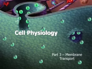
To permit survival and normal function of cultured tissues or cells several things have to be controlled: • pH • Temperature • Ionic strength • Access to essential nutrients • Animal and plant cells are put into nutrient-rich medium (culture medium) within specially coated plastic dishes or flasks
Culture medium often contains antibiotics • Cultures are manipulated in sterile cabinets (hoods) to prevent contamination from airborne microorganisms
Growing and Studying Cells in Culture • Animal cells grow in culture when supplied with necessary nutrients and appropriate 2D or 3D substrates. • Primary cells have a finite life span; transformed tumor cells and cell line cells can grow indefinitely. • A fluorescence-activated cell sorter (FACS) sorts labeled cells. • Hybridoma cells produce monoclonal antibodies that bind one antigen epitope and are used for basic research and therapeutics. • Basic biological processes can be studied by using genetic manipulation or drugs to interfere with specific cell components. • Large chemical libraries can be screened for compounds that target specific processes.
Media • Must supply 9 essential amino acids that cannot be synthesized by adult vertebrate cells • Phenylalanine • Valine • Threonine • Tryptophan • Isoleucine • Methionine • Leucine • Lysine • Histidine • Must also have 3 amino acids only produced by intact animal cells • Cysteine • Tyrosine • Arginine • Vitamins • Salts • Fatty acids • Glucose • Serum (proteins, insulin, growth factors, CAMs) • Other components depending on cell type
Primary Cell Line • Isolated directly from tissues • Normal tissues (liver, kidney, skin) or whole embryos • These cells have a finite number of times they will divide • Cease to grow after a time, senescence • Exception to this is the embryonic stem cell (ES)
Embryonic Stem Cells (ES) • Pluripotent – differentiate into wide range of cell types • Can be injected into the blastocoel of early mouse embryo and transplanted into uterus of pseudopregnantmouse • Induced Pluripotent Stem Cells (iPS) • Adult somatic cells that can be induced into pluripotency by exposing to certain proteins • Stem cell niches in multicellular organisms • Multipotent – give rise to some but NOT all cell types • Germ-line stems cells produce sperm and oocytes • Intestinal stem cells • Hematopoietic stem cells • Meristems in plants
Transformed Cell Line • Can grow indefinitely in culture • Often have abnormal DNA • Often have different chromosome #
Separating Cell Types • Some cells differ in density • Blood cells can be separated by centrifugation • Not all cells can be separated so easily so it must be done by other methods • Mark and sort • Flow cytometer • Fluorescence-activated cell sorter (FACS)
Differential labeling of cell types enables sorting from complex mixtures. Step 1: Labeled cells mixed with sheath fluid buffer solution pass single file through a laser light beam. Step 2: Both fluorescent light emitted and light scattered from each cell are measured and used to determine cell shape and size. Step 3: Forcing the cell suspension through a nozzle forms tiny droplets containing at most a single cell; each droplet obtains a negative electric charge proportional to fluorescence of that cell. Step 4: Passage through an electric field differentially deflects the droplets into different bins; as many as 10 million cells per hour can pass through the machine.
Growth of cells in Two-dimensional and three-dimensional culture mimics the in vivo environment
A wide variety of cell biological processes can be studied with cultured cells • Easily subjected to a variety of manipulations • Human diseases can be studied • New drugs can be tested • Old drugs – drugs that have been around for a long time but cell mechanism is not known; studying them can allow mapping out the exact effects on a particular cell type
Visualizing Cells • All microscopes produced magnified image of a small object (magnification) • Resolution – ability to distinguish between 2 closely positioned objects
In bright-field light microscopy, light from a tungsten lamp is focused on the specimen (usually stained with dyes to enhance contrast) by a condenser lens below the stage.
In phase-contrast (and differential-interference; DIC) microscopy , which increases contrast of biological specimens, incident light passing through an annular diaphragm focuses a circular annulus (ring) of light on the specimen. Light passing unobstructed through the specimen is focused by the objective lens onto the thicker gray ring of the phase plate, which absorbs some of the direct light and alters its phase by one-quarter of a wavelength. If a specimen refracts (bends) or diffracts the light because of the refractive index of the material, the phase of some light waves is altered (green lines), and the light waves pass through the clear region of the phase plate. The refracted and unrefracted light is recombined at the image plane to form the image. Click HERE to watch video
In fluorescence microscopy, a beam of light from a mercury lamp (gray lines) is directed to the excitation filter, which allows only the correct wavelength of light to pass through (green lines). The light is then reflected off a dichroic mirror and through the objective lens, which focuses it on the specimen. The fluorescent light emitted by the specimen (red lines) passes up through the objective lens, then through the dichroic mirror, and is focused and recorded on the detector at the image plane. Click HERE to watch video
Often, specimens for light microscopy are fixed • Solution often contains chemicals that cross-link most proteins and nucleic acids • Example is formaldehyde • After fixation, one of 2 things happens: • the specimen is embedded in paraffin and cut into sections • 50µm thick • Has to be done with a microtome • Cultured cells can be grown on coverslip and fixed and are thin enough that they don’t have to be sliced • Stain • Many chemical stains bind to molecules that have specific features • Examples: • Hematoxylin – binds to basic amino acids of certain proteins • Eosin – binds to acidic molecules (DNA, some amino acids)
What can fluorescence microscopy do? • Localize and quantify specific molecules in live cells • Quantify ion concentrations with ion-sensitive fluorescent dyes • Detect specific proteins in fixed cells
Green Fluorescent Protein – GFP • Jellyfish Aequorea Victoria • Other fluorescent proteins expressed by bacteria can be isolated for use
Conventional fluorescent microscopy has limitations • Out of focus fluorescent light • Deconvolution microscopy and Confocal Microscopy help to resolve this
Electron Microscopy • Electromagnetic lenses focus a high-velocity beam instead of visible light used by optical lenses • High resolution images • Single proteins, organelles, viruses can be imaged
Transmission Electron Microscopy • TEM samples are mounted on a small copper or • gold grid that is covered with a very thin film • of plastic and carbon to which a sample can adhere. • Specimens are stained with heavy metal, • such as uranyl acetate, and excess stain • is removed. • Negative stain of rotaviruses: The viruses • exclude the stain and are seen in negative outline.
Transmission Electron Microscopy • Metal Shadowing Poliovirus
Transmission Electron Microscopy • Specimens such as cells and tissues that are too thick have to fixed and sliced
Scanning Electron Microscopy • SEM allows visualization of the surfaces of metal coated items
Isolation of Cell Organelles • It is necessary to isolate organelles in their native state and identify and dissect the function of each component • Lysosomes, for example, had been identified by light microscopy • But their function was only known after a method was developed to isolate them • Isolation techniques: • Disruption of cells by vigorous homogenization, sonication, or other techniques releases their organelles. Swelling cells in a hypotonic solution weakens the plasma membrane, making it easier to rupture. • Sequential differential centrifugation of a cell homogenate yields fractions of organelles that differ in mass and density. • Equilibrium density-gradient centrifugation separates cellular components by density for purification. • Immunological techniques using antibodies against organelle- specific membrane proteins purify organelles and vesicles of similar sizes and densities.
What is the difference between a cell strain, a cell line and a clone? • A cell strain is a lineage of cells originating from a primary culture taken from an organism. Since these cells are not transformed, they have a limited lifespan in culture. • In contrast, a cell line is made of transformed cells and therefore these cells can divide indefinitely in culture. Such cells are said to be immortal. • A clone results when a single cell is cultured and gives rise to genetically identical progeny cells.