Download
1 / 1

E N D
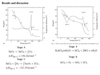
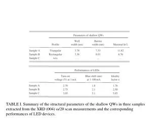
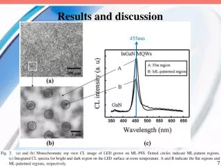
Abstract Scanning electron microscopy with X-ray micro-analysis combines the capability of elemental analysis with imaging (X-ray mapping). While elemental composition of geologic samples can be analyzed through destructive methods (atomic absorption spectroscopy), some are better left intact as their morphology provides clues that are lost when the sample is destroyed. Shape and composition of fossils combine to give clues concerning depositional environments and habitat at time of death. In this study, we analyze fossils recovered from Ordovician strata of the Dillsboro Formation (Madison Indiana). Specifically, we analyze the remains of an encrusting bryozoan colony on a brachiopod shell. Both of these marine animals have inorganic hard parts that have the potential to persist as fossils with or without mineral replacement. The original or possible replacement chemistry, and structure of these fossils is the emphasis of this study. Elemental analysis and X-ray mapping revealed that the hard parts of the organisms were similar in composition containing large amounts of calcium, as would be expected, and suggesting original shell composition. However, we found that the intrashell pore filling material of the bryozoan contains measurable amounts of aluminum, silicon, potassium, and iron, all absent or in much lower concentrations in the brachiopod remnants. This suggests infilling of primary porosity by siliciclastic mud matrix. Future work including quantitative analysis of thin cross sections of these fossils may improve our understanding of these organisms and their environment. Introduction Results and Discussion All fossils are preserved in sedimentary environments. Geologists use fossils to interpret depositional environments and to produce a biostratigraphic correlation within different rock units. Fossils are divided into body fossils and non-body fossils, also known as trace fossils (e.g. footprints). The unaltered remains of soft body parts are preserved by means of refrigeration, burial in bogs, or amber secretion. Hard body parts are also fossilized as unaltered remains, and are found as original bones, teeth, or shells of the organism. The altered remains of body fossils occur or form by replacement, recrystallization, compression, carbonization, and as molds and casts (Ref. 1). In this study, the specimen used is a body fossil; our interest is to gain insight into chemistry of its preservation through elemental analysis. The specimen is an encrusting bryozoan on a brachiopod shell. Geologic Strata of Interest The specimen was collected from the Dillsboro Formation of the Upper Ordovician strata in Southeastern Indiana off of U.S. Route 421, north of Madison, IN (figure 1A) from Indiana Geological Survey). The Dillsboro Formation is approximately 300 feet thick, and it overlies the Kope Formation and underlies the Saluda Formation. The formation consists of a richly fossiliferous series of alternating gray shales and thin slabs of coquina limestone, which are well-known to fossil collectors (Ref. 2). The fossils found in the Dillsboro Formation include brachiopods, bryozoans, molluscs, arthropods, and echinoderms. Fauna of the Ordovician Epeiric Sea By the Upper Ordovician (figure 1A), the continent was covered by a large shallow sea or epeiric sea, which deposited shale and limestone over most of the continental platform. The shallow, warm, calcium-carbonate rich environment was ideal for shell-secreting organisms. The most abundant organisms found in the Upper Ordovician, specifically the Dillsboro Formation, are bryozoans and brachiopods. Bryozoans found in the Upper Ordovician are marine animals. They are colonial organisms whose encrusting masses often contributed to the formation of limestones. An overview of the diversity of Ordovician Bryozoans can be seen in figure 2 (Ref. 4). In this study, the notable aspects of Bryozoan anatomy are their calcareous protective tube and the soft body it surrounds. Brachiopods are generally larger and more complex animals, their shells can be confused with those of bivalve molluscs. Brachiopod remains also contribute to the formation of limestone (Ref. 5). Elemental Analysis by Scanning Electron Microscopy (SEM) The electron beam formed by the SEM is composed of high energy electrons. When the electrons hit the sample, some are scattered back off the sample and allow us to form backscattered electron images (as seen in figure 3A-E). Beam electrons also cause specific energy transitions to occur in the specimen; these create X-rays whose energies are characteristic of specific elements. In order to get uncomplicated X-ray spectra, we imaged our samples without any conductive coating. This can lead to an unstable imaging condition called charging, which we eliminate by using the low vacuum mode of our SEM. This allows for a small amount of atmospheric moisture (water molecules) to enter the chamber and dissipate the electron charge. We collected X-rays generated by the sample with an energy dispersive spectrometer (EDS). This device is capable of measuring and counting X-ray photons; these counts are then plotted on an energy spectrum (figure 3F-I) or mapped to spatial coordinates (figure 4). All research was carried out using the environmental JEOL 5130 SEM with an Oxford Instruments Pentafet thin window EDS. This detector has a beryllium (Be) window which can detect elements with atomic numbers > 10. The microscope has a tungsten filament, we used the solid state backscattered electron detector. F. A. B. Elemental Analysis by SEM of Late Ordovician Brachiopod and Bryozoan FossilsWonnell, Chloe and Neff, David. Department of Geology, College of Science, Marshall University G. C. D. Y-axis units are # X-ray photon counts H. Figure 3: Backscattered electron (BSE) images taken in low vacuum mode to prevent specimen charging (A-E) and X-ray spectra (F-I). Brachiopod (A, B, and F), bryozoan (C, D, G, and H), and in E, a second smaller brachiopod shell (arrow) overlying encrusting bryozoan. 3D is an image of intrashell area of the Bryozoan, the cube shaped crystal in D. has higher relative Mg than the sur-rounding material (data for crystal not shown). Spectrum I. is from the rock as seen in figure 2B, left. All X-ray spectra are qualitative, scale is normalized to the largest calcium Kα peak. E. I. B. A. A. From the context of the collection site, we know the age of the source strata to be Upper Ordovician (see introduction). The animals whose remains are shown here are known to have lived in a shallow epeiric sea that once covered much of the continent. Consistent with that habitat are the calcareous remains of the hard parts of the animals. This type of anatomical adaptation, calcium rich shell, is very common in former and present shallow sea dwellers. Both animals seen in this study were dominant in the shallow seas of the Paleozoic, but have now been supplanted by other calcium fixing organisms such as mollusks (Ref. 6). The X-ray spectra in figure 3 F, G, and H and the X-ray maps in figure 4 show that shells (also known as carbonate skeleton) of both brachiopod and bryozoan are rich in calcium, presumably some form of calcite. Whether the ratios of elements seen in the spectra represent the original composition of the shells or replacement minerals cannot be determined from this data. In contrast, the pore filling intrashell material of the bryozoan is identified by the silicon, aluminum, and magnesium peaks (figure 3H), and in the corresponding X-ray map (figure 4A and C). The finding of these elements in the intrashell regions of the bryozoan is of interest, because it implies replacement by elements other than calcium, which is found predominately in the outer tubing of the bryozoan and brachiopod shell (figure 4 A and C). It was suggested (by personal correspondence, Dr. Martino) that these elements indicate filling by a mud matrix. This finding is consistent with the Dillsboro Formation (see introduction) that has a lithology of mostly coquina limestone and gray shales (Ref. 7). The rock spectrum (figure 3 I) was taken from the limestone matrix that surrounded the fossil as it was in situ. We think silicate minerals related to the gray shale may have filled the intrashell areas (formerly occupied by the animals’ soft body) of the bryozoan. The dominance of calcium and the absence of magnesium in the brachiopod shell (figure 3A, B, F) helped us reason that the brachiopod shell was likely replaced by low-Mg calcite during diagenesis. In studies of calcium carbonate minerals, specifically of low-Mg calcite and high-Mg calcite, it is known that carbonate sediments are often converted to low-Mg calcite, and that high-Mg calcite at times will lose its Mg (Ref. 6). Based on this knowledge, we hypothesize that our fossilized brachiopod may have undergone diagenesis from high-Mg calcite to low-Mg-calcite. Future work may involve this hypothesis by analyzing cross sections. X-axis units are X-ray photon energy A. B. C. http://geology.com/time/geologic-time-scale-550.gif Figure 4: X-ray maps of the fossil show spatial distribution of detected elements. A is bryozoan, B is brachiopod. C is both with bryozoan at left side of each pane and brachiopod at lower right of each pane. Elements mapped in each pane are identified at top of pane, the 5310lv pane is a BSE image. Figure 1: Above left (A) is a general stratigraphic column of Paleozoic rocks in Indiana, the red box indicates the formation of origin for the fossils in this study. At right (B) we see where the Ordovician period falls in the overall geologic time scale. Figure 2A: The SEM images above, from reference 4 show the diversity of Ordovician bryozoans. These specimens were collected from an other Ordovician epeiric sea in present day India. Scale bars represent 0.5 mm in 1–2, 4–8, 10, 12, 15, 17; 1 mm in 3, 9, 11, 13–14, 16; 2 mm in 18. Figure 2B: A photograph of the back of specimen showing the rock matrix from which the fossil was collected (left). The photograph at right shows the brachiopod shell (approximately 1.5 inches across). The white arrow points to the encrusting bryozoan. References B. 1. Pullen, Stephanie. “What is a fossil?” UCMP. April 2004. 20 March 2009. <http://www.ucmp.berkeley.edu/paleo/fossils/index.html>.2. Wahlman, G.P., 1992, Middle and Upper Ordovician symmetrical univalved mollusks (Monoplacophora and Bellerophontina) of the Cincinnati arch region, IN Pojeta, John, Jr., ed., Contributions to the Ordovician paleontology of Kentucky and nearby states: U.S. Geological Survey Professional Paper, 1066-O, p. O1-O203.3. “Geologic Time Scale.” Geology.com . 2005. 12 April 2009. <http://geology.com/time/geologic-time-scale-550.gif>.4. Suttner, Thomas J., and Ernst, Andrej. “Upper Ordovician Bryozoans of the Pin Formation (Spiti Valley, Northern India).” Paleontology. 50 (2007): 1485-1518.5. Cooper, John D., Miller, Richard H., and Patterson, Jacqueline. A Trip Through Time: Principles of Historical Geology. 2nd ed. Columbus: Merrill, 1990.6. Tucker, Maurice E. Sedimentary Petrology: An Introduction to the Origin of Sedimentary Rocks. 3rd ed. Alden Press: Blackwell Science, 2001. 7. Bevis, Kenneth, Worcester, Pete, Totten, Stanley M. Geology Laboratory Manual. 10th ed. Hanover: Hanover College, 2007. Acknowledgements Thanks to Dr. Martino and Dr. El-Shazly for their advice. And thanks to Marshall University‘s MBIC for maintenance of SEM facilities. http://www.marshall.edu/mbic/