Download
1 / 60
620 likes | 1.1k Vues
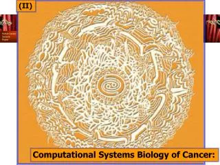
(I). Computational Systems Biology of Cancer:. Professor of Computer Science, Mathematics and Cell Biology ¦ Courant Institute, NYU School of Medicine, Tata Institute of Fundamental Research, and Mt. Sinai School of Medicine. Bud Mishra. War on Cancer.

E N D
(I) Computational Systems Biology of Cancer:
Professor of Computer Science, Mathematics and Cell Biology ¦ Courant Institute, NYU School of Medicine, Tata Institute of Fundamental Research, and Mt. Sinai School of Medicine Bud Mishra
War on Cancer • “Reports that say that something hasn't happened are always interesting to me, because as we know, there are known knowns; there are things we know we know. We also know there are known unknowns; that is to say we know there are some things we do not know. But there are also unknown unknowns – the ones we don't know we don't know.” • US Secretary of Defense, Mr. Donald Rumsfeld, Quoted completely out of context.
Introduction: Cancer and Genomics: What we know & what we do not “Cancer is a disease of the genome.”
Outline • Genomics • Genome Modification & Repair • Segmental Duplications Models
Genomics • Genome: • Hereditary information of an organism is encoded in its DNA and enclosed in a cell (unless it is a virus). All the information contained in the DNA of a single organism is its genome. • DNA molecule can be thought of as a very long sequence of nucleotides or bases: S = {A, T, C, G}
Complementarity • DNA is a double-stranded polymer and should be thought of as a pair of sequences over S. • However, there is a relation of complementarity between the two sequences: • A , T, C , G
DNA Structure. • The four nitrogenous bases of DNA are arranged along the sugar- phosphate backbone in a particular order (the DNA sequence), encoding all genetic instructions for an organism. • Adenine (A) pairs with thymine (T), while cytosine (C) pairs with guanine (G). • The two DNA strands are held together by weak bonds between the bases.
Structure and Components • Complementary base pairs (A-T and C-G) • Cytosine and thymine are smaller (lighter) molecules, called pyrimidines • Guanine and adenine are bigger (bulkier) molecules, called purines. • Adenine and thymine allow only for double hydrogen bonding, while cytosine and guanine allow for triple hydrogen bonding.
Inert & Rigid • Thus the chemical (hydrogen bonding) and the mechanical (purine to pyrimidine) constraints on the pairing lead to the complementarity and makes the double stranded DNA both chemically inert and mechanically quite rigid and stable.
Transcription Translation DNA RNA Protein The Central Dogma • The central dogma(due to Francis Crick in 1958) states that these information flows are all unidirectional: “The central dogma states that once `information' has passed into protein it cannot get out again.”
Transcription Translation DNA RNA Protein The Central Dogma “…The transfer of information from nucleic acid to nucleic acid, or from nucleic acid to protein, may be possible, but transfer from protein to protein, or from protein to nucleic acid is impossible. Information means here the precise determination of sequence, either of bases in the nucleic acid or of amino acid residues in the protein.”
DNA RNA Protein Genome Evolution Phenotype Selection Genotype Transcription Translation Part-lists, Annotation, Ontologies The New Synthesis
Cancer Initiation and Progression Mutations, Translocations, Amplifications, Deletions Epigenomics (Hyper & Hypo-Methylation) Alternate Splicing Cancer Initiation and Progression Proliferation, Motility, Immortality, Metastasis, Signaling
Multi-step Nature of Cancer: • Cancer is a stepwise process, typically requiring accumulation of mutations in a number of genes. • ~6-7 independent mutations typically occur over several decades: • Conversion of proto-oncogenes to oncogenes • Inactivation of tumor suppressor gene
Amplifications & Deletions Mutation in a TSG Epigenomics Conversion of a Proto-Oncogene Deletion of a TSG Deletion of a TSG
The Cancer Genome Atlas • Obtain a comprehensive description of the genetic basis of human cancer. • Identify and characterize all the sites of genomic alteration associated at significant frequency with all major types of cancers.
The Cancer Genome Atlas • Increase the effectiveness of research to understand • tumor initiation and progression, • susceptibility to carcinogensis, • development of cancer therapeutics, • approaches for early detection of tumors & • the design of clinical trials.
Specific Goals • Identify all genomic alterations significantly associated with all major cancer types. • Such knowledge will propel work by thousands of investigators in cancer biology, epidemiology, diagnostics and therapeutics.
Create large collection of appropriate, clinically annotated samples from all major types of cancer; and Characterize each sample in terms of: All regions of genomic loss or amplification, All mutations in the coding regions of all human genes, All chromosomal rearrangements, All regions of aberrant methylation, and Complete gene expression profile, as well as other appropriate technologies. To Achieve this goal …
Biomedical Rationale • Cancer is a heterogeneous collection of heterogeneous diseases. • For example, prostate cancer can be an indolent disease remaining dormant throughout life or an aggressive disease leading to death. • However, we have no clear understanding of why such tumors differ.
Biomedical Rationale • Cancer is fundamentally a disease of genomic alteration. • Cancer cells typically carry many genomic alterations that confer on tumors their distinctive abilities (such as the capacity to proliferate and metastasize, ignoring the normal signals that block cellular growth and migration) and liabilities (such as unique dependence on certain cellular pathways, which potentially render them sensitive to certain treatments that spare normal cells).
History • 1960s • The genetic basis of cancer was clear from cytogenetic studies that showed consistent translocations associated with specific cancers (notably the so-called Philadelphia chromosome in chronic myelogenous leukemia). • 1970s • Recognize specific cancer-causing mutations through recombinant DNA revolution of the 1970s. • The identification of the first vertebrate and human oncogenes and the first tumor suppressor genes, • These discoveries have elucidated the cellular pathways governing processes such as cell-cycle progression, cell-death control, signal transduction, cell migration, protein translation, protein degradation and transcription. • For no human cancer do we have a comprehensive understanding of the events required.
Gene resequencing. Specific gene classes (such as kinases and phosphatases) in particular cancer types. Epigenetic changes. Loss of function of tumor suppressor genes by epigenetic modification of the genome — such as DNA methylation and histone modification. Genomic loss and amplification. Consistent association with genomic loss or amplification in many specific regions, indicating that these regions harbor key cancer associated genes Chromosome rearrangements. Activate kinase pathways through fusion proteins or inactivating differentiation programs through gene disruption. Hematological malignancies: a single stereotypical translocation in some diseases (such as CML) and as many as 20 important translocations in others (such as AML). Adult solid tumors have not been as well characterized, in part owing to technical hurdles. Scientific Foundation for a Human Cancer Genome Project
EBD • J.B.S. Haldane (1932): • “A redundant duplicate of a gene may acquire divergent mutations and eventually emerge as a new gene.” • Susumu Ohno (1970): • “Natural selection merely modified, while redundancy created.”
UTP UMP UDP Organism CTP Living cells Leu3 Functional modules BAT1 LEU1 ILV2 Motifs in cellular etworks Metabolic pathways Regulatory motifs Interaction clusters Non-random distribution in Genomic data ? Short words frequency Protein family size Long range orrelation Genome Evolution Evolution by Duplication
Mer-scape… • Overlapping words of different sizes are analyzed for their frequencies along the whole human genome • Red: 24-mers, • Green: 21-mers • Blue:18 mers • Gray:15 mers • To the very left is a ubiquitous human transposon Alu. The high frequency is indicative of its repetitive nature. • To the very right is the beginning of a gene. The low frequency is indicative of its uniqueness in the whole genome.
Doublet Repeats • Serendipitous discovery of a new uncataloged class of short duplicate sequences; doublet repeats. • almost always < 100 bp • (Top) . The distance between the two loci of a doublet is plotted versus the chromosomal position of the first locus. • (Bottom) : Distribution of doublets (black) and segmental duplications (red) across human chromosome 2
Segmental Duplications • 3.5% ~ 5% of the human genome is found to contain • segmental duplications, with length > 5 or 1kb, identity > 90%. • These duplications are estimated to have emerged about 40Mya under neutral assumption. • The duplications are mostly interspersed (non-tandem), and happen both inter- and intra-chromosomally. Human From [Bailey, et al. 2002]
f - - f - - deletion or mutation insertion f + - f + - Duplication by recombination between other repeats or other mechanisms deletion or mutation insertion f ++ f ++ Duplication by recombination between repeats Mutation accumulation in the duplicated sequences The Model
Time after duplication 1-α-2β 1-α-2β 1-α-2β h0-- α α α α f - - h1-- γ 2β γ 2β 2β γ h0+- h0 α α α α H0 f + - 1-α-β/2-γ 1-α-β/2-γ 1-α-β/2-γ 2γ β/2 2γ β/2 2γ β/2 h0++ α α α α H1 f ++ h1 h1++ 1-α-2γ 1-α-2γ 1-α-2γ The Mathematical Model 0 ≤ d < ε ε ≤ d < 2ε (k-1)ε ≤ d < kε h1: proportion of duplications by repeat recombination; h1++: proportion of duplications by recombination of the specific repeat; h1- -: proportion of duplications by recombination of other repeats; h0: proportion of duplications by other repeat-unrelated mechanism; h0++: proportion of h0 with common specific repeat in the flanking regions; h0+-: proportion of h0 with no common specific repeat in the flanking regions; h0- -: proportion of h0 with no specific repeat in the flanking regions; α: mutation rate in duplicated sequences; β: insertion rate of the specific repeat; γ: mutation rate in the specific repeat; d: divergence level of duplications; ε: divergence interval of duplications.
Alu L1 f - - f - - f + - f + - f ++ f ++ Diversity: Diversity: Model Fitting • The model parameters (αAlu, βAlu, γAlu, αL1, βL1, γL1) are estimated from the reported mutation and insertion rates in the literature. • The relative strengths of the alternative hypotheses can be estimated by model fitting to the real data. • h1Alu ≈ 0.76; h1++Alu≈0.3; h1L1 ≈ 0.76; h1++L1≈0.35.
Random drawn Put back Duplication Polya’s Urn
F1 (decide an initial position) F3 (decide copy number) F2 (decide selected length) k copies F4 (decide insertion positions) Repetitive Random Eccentric GOD • Genome Organizing Devices (GOD) • Polya’s Urn Model: • F’s: functions deciding probability distributions
Insertion (duplication): Deletion: DNA polymerase stuttering(replication slippage) normal replication DNA polymerase normal replication polymerase pausing and dissociation polymerase pausing and dissociation 3’ realignment and polymerase reloading 3’ realignment and polymerase reloading
Transposons~ causes: deletions and duplications • Transposon actions in • genomic DNA: Donor DNA • Duplication: transposon DNA intermediates Target DNA Transposase cuts in target DNA • Deletion: transposon IS IS Transposon looping out Transposon inserted Transposon deleted DNA is repaired-resulting in a duplication of the transposon and target site
MSH6 MSH2 Exo MSH2 MSH6 MSH2 MSH6 PMS2 MLH1 MLH1 PMS2 DNA mismatch repair mechanism~ prevents duplications and deletions Mismatch recognition on daughter strand Degradation of the mismatched daughter strand in the a-loop DNA a-loop formation by translocation through the proteins Refilling the gap by DNA polymerase Corrected daughter strand DNA polymerase
Graph Model A. Deletion: With probability p0 • Graph description • G = {V, E}, G is a directed multi-graph; • V {Vi, all n-mer’s, i = 1…4n}, ; • E { (Vi , Vj ), when Vi represents the n-mer immdiately upstream of the n-mer represented by Vj in the genomic sequence}; • ki = incoming (or outgoing) degree of node i (Vi) = copy number of the n-mer represented by Vi; • During the graph evolution, at each iteration, one of the following happens: deletion (with probability p0), duplication (with probability p1) or substitution (with probability q). p0 + p1 + q = 1. • For an arbitrary node Vi , the probabilities of one of the above events happens is as follows: B. Duplication:With probability p1 C. Substitution: With probability q
Real distribution from genome analysis Expected distribution from random sequences Model fitting results Model Fitting
Model Fitted Parameter • The substitution rate, q, increases with the sizes of mer’s. • The ratio between duplication and deletion rate, p1/p0, increases with sizes of mer’s. • The substitution rate, q, tends to decrease when the genome sizes are larger. Especially, q is much smaller in eukaryotic genomes than in prokaryotic genomes.
J.B.S. Haldane • “If I were compelled to give my own appreciation of the evolutionary process…, I should say this: In the first place it is very beautiful. In that beauty, there is an element of tragedy…In an evolutionary line rising from simplicity to complexity, then often falling back to an apparently primitive condition before its end, we perceive an artistic unity … • “To me at least the beauty of evolution is far more striking than its purpose.” • J.B.S. Haldane, The Causes of Evolution. 1932.
Normal epithelial mucosa Neoplastic polyp Cancer
A Challenge • “At present, description of a recently diagnosed tumor in terms of its underlying genetic lesions remains a distant prospect. Nonetheless, we look ahead 10 or 20 years to the time when the diagnosis of all somatically acquired lesions present in a tumor cell genome will become a routine procedure.” • Douglas Hanahan and Robert Weinberg • Cell, Vol. 100, 57-70, 7 Jan 2000
CGH:Comparative Genomic Hybridization. • Equal amounts of biotin-labeled tumor DNA and digoxigenin-labeled normal reference DNA are hybridized to normal metaphase chromosomes • The tumor DNA is visualized with fluorescein and the normal DNA with rhodamine • The signal intensities of the different fluorochromes are quantitated along the single chromosomes • The over-and underrepresented DNA segments are quantified by computation of tumor/normal ratio images and average ratio profiles Amplification Deletion