Chapter 47
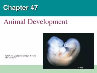
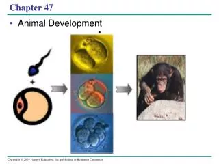
Chapter 47. Animal Development. Overview: A Body-Building Plan. It is difficult to imagine that each of us began life as a single cell ( fertilized egg ) called a zygote . A human embryo at about 6–8 weeks after conception shows development of distinctive features.

Chapter 47
E N D
Presentation Transcript
Chapter 47 Animal Development
Overview: A Body-Building Plan • It is difficult to imagine that each of us began life as a single cell (fertilized egg) called a zygote. • A human embryo at about 6–8 weeks after conception shows development of distinctive features.
How did this complex embryo develop from a single fertilized egg? 1 mm
Development is determined by the zygote’s genome and molecules in the egg cytoplasm called Cytoplasmic determinants. • Cell differentiationis the specialization of cells in structure and function. • Morphogenesisis the process by which an animal takes shape / form. • Model organisms are species that are representative of a larger group and easily studied. Classic embryological studies use the sea urchin, frog, chick, and the nematode C. elegans.
After fertilization, embryonic development proceeds through cleavage, gastrulation, and organogenesis • Important events regulating development occur during fertilization and the three stages that build the animal’s body • Cleavage: cell division creates a hollow ball of cells called a blastula • Gastrulation: cells are rearranged into a three-layered gastrula • Organogenesis: the three germ layers interact and move to give rise to organs.
Fertilization: sperm + egg = zygoten + n = 2n • Fertilization brings the haploid nuclei of sperm and egg together, forming a diploid zygote. • The sperm’s contact with the egg’s surface initiates metabolic reactions in the egg that trigger the onset of embryonic development: • Acrosomal Reaction • Cortical Reaction
The Acrosomal Reaction • The acrosomal reactionis triggered when the sperm meets the egg. • The acrosome at the tip of the sperm releases hydrolytic enzymes that digest material surrounding the egg. • Gamete contact and/or fusion depolarizes the egg cell membrane and sets up a fast block to polyspermy.
Theacrosomaland cortical reactions during sea urchin fertilization Sperm plasmamembrane Spermnucleus Fertilizationenvelope Acrosomalprocess Basal body(centriole) Actinfilament Spermhead Corticalgranule Fusedplasmamembranes Perivitellinespace Hydrolytic enzymes Acrosome Jelly coat Vitelline layer Sperm-bindingreceptors Egg plasmamembrane EGG CYTOPLASM
The Cortical Reaction • Fusion of egg and sperm also initiates the cortical reaction: • This reaction induces a rise in Ca2+ that stimulates cortical granulesto release their contents outside the egg. • These changes cause formation of a fertilization envelopethat functions as a slow block to polyspermy.
Activation of the Egg • The sharp rise in Ca2+ in the egg’s cytosol increases the rates of cellular respiration and protein synthesis by the egg cell. • With these rapid changes in metabolism, the egg is said to be activated. • The sperm nucleus merges with the egg nucleus and cell division begins.
Fertilization in Mammals • Fertilization in mammals and other terrestrial animals is internal. • In mammalian fertilization, the cortical reaction modifies the zona pellucida,the extracellular matrix of the egg,as a slow block to polyspermy. • In mammals the first cell division occurs 12–36 hours after sperm binding. • The diploid nucleus forms after this first division of the zygote.
Fertilization in mammals Zona pellucida Follicle cell Cortical granules Spermnucleus Spermbasal body
Cleavage = Rapid Mitosis / No Mass change • Fertilization is followed by cleavage, a period of rapid cell division without growth. • Cleavage partitions the cytoplasm of one large cell into many smaller cells called blastomeres. • Theblastula is a ball of cells with a fluid-filled cavity called a blastocoel.
Cleavage in an echinoderm embryo (a) Fertilized egg (b) Four-cell stage (c) Early blastula (d) Later blastula
The eggs and zygotes of many animals, except mammals, have a definite polarity. • The polarity is defined by distribution of yolk(stored nutrients). • The vegetal pole has more yolk; the animal pole has less yolk.
The three body axes are established by the egg’s polarity and by a cortical rotation following binding of the sperm. • Cortical rotation exposes a gray crescent opposite to the point of sperm entry.
Dorsal The body axes and their establishment in an amphibian Right Anterior Posterior (a) The three axes of the fully developed embryo Left Ventral Animal pole Firstcleavage Pigmentedcortex Point ofspermnucleusentry Animalhemisphere Futuredorsalside Vegetalhemisphere Graycrescent Vegetal pole - yolk (b)Establishing the axes
Cleavage planes usually follow a pattern that is relative to the zygote’s animal and vegetal poles. • Cell division is slowed by yolk. Yolk can cause uneven cell division at the poles. • Holoblastic cleavage, complete division of the egg, occurs in species whose eggs have little or moderate amounts of yolk, such as sea urchins and frogs. • Meroblastic cleavage, incomplete division of the egg, occurs in species with yolk-rich eggs, such as reptiles and birds.
Cleavagein a frog embryo 0.25 mm 0.25 mm Animal pole Blastocoel Vegetalpole: yolk Zygote 2-cellstageforming 4-cellstageforming 8-cellstage Blastula(crosssection)
Gastrulation • Gastrulation rearranges the cells of a blastula into a three-layered embryo, called a gastrula, which has a primitive gut. • The three layers produced by gastrulation are called embryonic germ layers: • The ectoderm forms the outer layer • The endodermlines the digestive tract • The mesoderm partly fills the space between the endoderm and ectoderm.
Gastrulation in the sea urchin embryo: The blastula consists of a single layer of cells surrounding the blastocoel. Mesenchyme cells migrate from the vegetal pole into the blastocoel. The vegetal plate forms from the remaining cells of the vegetal pole and buckles inward through invagination. The newly formed cavity is called the archenteron. This opens through the blastopore, which will become the anus.
Gastrulation in a sea urchin embryo Key Future ectoderm Future mesoderm Future endoderm Vegetal Pole Invagination Blastocoel Filopodiapullingarchenterontip Animalpole Archenteron - cavity Blastocoel Blastocoel Blastopore Mesenchymecells Ectoderm Vegetalplate Vegetalpole Mouth Mesenchymecells Mesenchyme(mesodermforms futureskeleton) Digestive tube (endoderm) Blastopore 50 µm Anus (from blastopore)
Gastrulation in the frog • The frog blastula is many cell layers thick. Cells of the dorsal liporiginate in the gray crescentand invaginate to create the archenteron. • Cells continue to move from the embryo surface into the embryo by involution. These cells become the endoderm and mesoderm. • The blastopore encircles a yolk plug when gastrulation is completed. • The surface of the embryo is now ectoderm, the innermost layer is endoderm, and the middle layer is mesoderm.
Gastrulation in a frog embryo SURFACE VIEW CROSS SECTION Animal pole Blastocoel Dorsal lipof blasto-pore Dorsal lipof blastopore Blastopore Earlygastrula Vegetal pole Blastocoelshrinking Archenteron Ectoderm Mesoderm Blastocoelremnant Endoderm Archenteron Key Blastopore Future ectoderm Lategastrula Future mesoderm Yolk plug Blastopore Future endoderm
Gastrulation in the chick • The embryo forms from a blastoderm and sits on top of a large yolk mass. • During gastrulation, the upper layer of the blastoderm (epiblast) moves toward the midline of the blastoderm and then into the embryo toward the yolk. • The midline thickens and is called the primitive streak. • The movement of different epiblast cells gives rise to the endoderm, mesoderm, and ectoderm.
Gastrulation in a chick embryo Dorsal Fertilized egg Primitive streak Anterior Embryo Left Right Yolk Posterior Ventral Primitive streak Epiblast Futureectoderm Blastocoel Endoderm Migratingcells(mesoderm) Hypoblast YOLK
Organogenesis • During organogenesis, various regions of the germ layers develop into rudimentary organs. • The frog is used as a model for organogenesis. • Early in vertebrate organogenesis, the notochordforms from mesoderm, and the neural plate forms from ectoderm.
Early organogenesis in a frog embryo Eye Somites Tail bud Neural folds Neural plate Neuralfold SEM 1 mm 1 mm Neural crestcells Neural tube Neuralfold Neural plate Notochord Coelom Neural crestcells Somite Notochord Ectoderm Archenteron(digestivecavity) Outer layerof ectoderm Mesoderm Endoderm Neural crestcells (c) Somites Archenteron (a) Neural plate formation Neural tube (b) Neural tube formation
The neural plate soon curves inward, forming the neural tube. The neural tube will become the central nervous system = brain and spinal cord. • Neural crest cells develop along the neural tube of vertebrates and form various parts of the embryo: nerves, parts of teeth, skull bones ... • Mesoderm lateral to the notochord forms blocks called somites. • Lateral to the somites, the mesoderm splits to form the coelom.
Organogenesis in a chick embryo is similar to that in a frog Eye Neural tube Notochord Forebrain Somite Heart Coelom Archenteron Endoderm Lateral fold Mesoderm Bloodvessels Ectoderm Somites Yolk stalk Yolk sac These layersform extraembryonicmembranes Neural tube YOLK (a) Early organogenesis (b) Late organogenesis
Adult derivatives of the three embryonic germ layers in vertebrates ECTODERM MESODERM ENDODERM NotochordSkeletal systemMuscular systemMuscular layer ofstomach and intestineExcretory systemCirculatory and lymphaticsystems Reproductive system(except germ cells) Dermis of skinLining of body cavityAdrenal cortex Epidermis of skin and itsderivatives (including sweatglands, hair follicles)Epithelial lining of mouthand anusCornea and lens of eyeNervous systemSensory receptors inepidermisAdrenal medullaTooth enamelEpithelium of pineal andpituitary glands Epithelial lining ofdigestive tractEpithelial lining ofrespiratory systemLining of urethra, urinarybladder, and reproductivesystemLiverPancreasThymusThyroid and parathyroidglands
Developmental Adaptations of Amniotes • Embryos of birds, other reptiles, and mammals develop in a fluid-filled sac in a shell or the uterus. • Organisms with these adaptations are called amniotes. • Amniotes develop extra-embryonic membranes to support the embryo.
Amniote ExtraEmbryonic Membranes • During amniote development, four extraembryonic membranes form around the embryo: • The chorionoutermost membrane / functions in gas exchange. • The amnionencloses the amniotic fluid. • The yolk sacencloses the yolk. • The allantoisdisposes of nitrogenous waste products and contributes to gas exchange.
ExtraEmbryonic Membranes in birds and other reptiles: Amnion Allantois Embryo Amnioticcavitywithamniotic fluid Albumen Shell Yolk (nutrients) Chorion Yolk sac
Mammalian Development • The eggs of placental mammals • Are small yolk and store few nutrients • Exhibit holoblastic cleavage • Show no obvious polarity. • Gastrulation and organogenesis resemble the processes in birds and other reptiles. • Early cleavage is relatively slow in humans and other mammals.
At completion of cleavage, the blastocystforms. • A group of cells called the inner cell mass develops into the embryo and forms the extraembryonic membranes. • The trophoblast, the outer epithelium of the blastocyst, initiates implantation in the uterus, and the inner cell mass of the blastocyst forms a flat disk of cells. • As implantation is completed, gastrulation begins.
Early embryonic development of a human Endometrialepithelium(uterine lining) Uterus Inner cell mass Trophoblast Blastocoel
Early embryonic development of a human Expandingregion oftrophoblast Maternalbloodvessel Epiblast Hypoblast Trophoblast
The epiblast cells invaginate through a primitive streak to form mesoderm and endoderm. • The placenta is formed from the trophoblast, mesodermal cells from the epiblast, and adjacent endometrial tissue. • The placenta allows for the exchange of materials between the mother and embryo. • By the end of gastrulation, the embryonic germ layers have formed. The extraembryonic membranes in mammals are homologous to those of birds and other reptiles and develop in a similar way.
Early embryonic development of a human Expandingregion oftrophoblast Amnioticcavity Epiblast Hypoblast Yolk sac (fromhypoblast) Extraembryonicmesoderm cells(from epiblast) Chorion (fromtrophoblast)
Early embryonic development of a human Amnion Chorion Ectoderm Mesoderm Endoderm Yolk sac Extraembryonicmesoderm Atlantois
Four stages in early embryonic development of a human Endometrialepithelium(uterine lining) Expandingregion oftrophoblast Maternalbloodvessel Uterus Inner cell mass Epiblast Trophoblast Hypoblast Blastocoel Trophoblast Expandingregion oftrophoblast Amnion Amnioticcavity Chorion Ectoderm Epiblast Mesoderm Hypoblast Endoderm Yolk sac (fromhypoblast) Yolk sac Extraembryonicmesoderm cells(from epiblast) Extraembryonicmesoderm Chorion (fromtrophoblast) Allantois
Morphogenesisin animals involves specific changes in cell shape, position,and adhesion • Morphogenesis is a major aspect of development in plants and animals. • Only in animals does it involve the movement of cells.
The Cytoskeleton, Cell Motility, and Convergent Extension • Changes in cell shape usually involve reorganization of the cytoskeleton. • Microtubules and microfilaments affect formation of the neural tube.
Change in cell shape during morphogenesis Ectoderm Neuralplate Microtubules Actin filaments Neural tube
The cytoskeleton also drives cell migration, or cell crawling, the active movement of cells. • In gastrulation, tissue invagination is caused by changes in cell shape and migration. • Cell crawling is involved in convergent extension, a morphogenetic movement in which cells of a tissue become narrower and longer.
Role of Cell Adhesion Molecules and the Extracellular Matrix • Cell adhesion molecules located on cell surfaces contribute to cell migration and stable tissue structure. • One class of cell-to-cell adhesion molecule is the cadherins, which are important in formation of the frog blastula.
Cadherin is required for development of the blastula RESULTS 0.25 mm 0.25 mm Control embryo Embryo without EP cadherin
The developmental fate of cells depends on their history and on inductive signals • Cells in a multicellular organism share the same genome. • Differences in cell types is the result of differentiation, the expression of different genes = differential gene expression.
Two general principles underlie differentiation 1. During early cleavage divisions, embryonic cells must become different from one another. • If the egg’scytoplasm is heterogenous, dividing cells vary in thecytoplasmic determinantsthey contain. 2. After cell asymmetries are set up, interactions among embryonic cells influence their fate, usually causing changes in gene expression • This mechanism is calledinduction, and is mediated by diffusible chemicals or cell-cell interactions.