Animal Development Overview: The Body-Building Plan
Explore the journey from zygote to animal, focusing on cleavage, gastrulation, and organogenesis, including fertilization processes and key developmental events.

Animal Development Overview: The Body-Building Plan
E N D
Presentation Transcript
Chapter 47 Animal Development
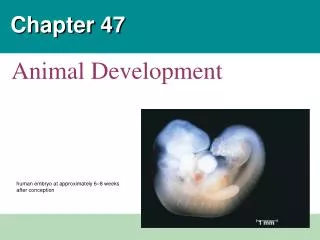
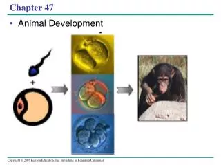
Overview: A Body-Building Plan • It is difficult to imagine that each of us began life as a single cell called a zygote • A human embryo at about 6–8 weeks after conception shows development of distinctive features
Fig. 47-1 1 mm
The question of how a zygote becomes an animal has been asked for centuries • As recently as the 18th century, the prevailing theory was called preformation • Preformation is the idea that the egg or sperm contains a miniature infant, or “homunculus,” which becomes larger during development
Development is determined by the zygote’s genome and molecules in the egg called cytoplasmic determinants • Cell differentiation is the specialization of cells in structure and function • Morphogenesis is the process by which an animal takes shape
Model organisms are species that are representative of a larger group and easily studied, for example, Drosophila and Caenorhabditis elegans • Classic embryological studies have focused on the sea urchin, frog, chick, and the nematode C. elegans
Concept 47.1: After fertilization, embryonic development proceeds through cleavage, gastrulation, and organogenesis • Important events regulating development occur during fertilization and the three stages that build the animal’s body • Cleavage: cell division creates a hollow ball of cells called a blastula • Gastrulation: cells are rearranged into a three-layered gastrula • Organogenesis: the three layers interact and move to give rise to organs
Fertilization • Fertilization brings the haploid nuclei of sperm and egg together, forming a diploid zygote • The sperm’s contact with the egg’s surface initiates metabolic reactions in the egg that trigger the onset of embryonic development
The Acrosomal Reaction • The acrosomal reaction is triggered when the sperm meets the egg • The acrosome at the tip of the sperm releases hydrolytic enzymes that digest material surrounding the egg
Fig. 47-3-1 Basal body(centriole) Spermhead Acrosome Jelly coat Vitelline layer Sperm-bindingreceptors Egg plasmamembrane
Fig. 47-3-2 Basal body(centriole) Spermhead Hydrolytic enzymes Acrosome Jelly coat Vitelline layer Sperm-bindingreceptors Egg plasmamembrane
Fig. 47-3-3 Spermnucleus Acrosomalprocess Basal body(centriole) Actinfilament Spermhead Hydrolytic enzymes Acrosome Jelly coat Vitelline layer Sperm-bindingreceptors Egg plasmamembrane
Fig. 47-3-4 Sperm plasmamembrane Spermnucleus Acrosomalprocess Basal body(centriole) Actinfilament Spermhead Fusedplasmamembranes Hydrolytic enzymes Acrosome Jelly coat Vitelline layer Sperm-bindingreceptors Egg plasmamembrane
Fig. 47-3-5 Sperm plasmamembrane Spermnucleus Fertilizationenvelope Acrosomalprocess Basal body(centriole) Actinfilament Spermhead Corticalgranule Fusedplasmamembranes Perivitellinespace Hydrolytic enzymes Acrosome Jelly coat Vitelline layer Sperm-bindingreceptors Egg plasmamembrane EGG CYTOPLASM
Gamete contact and/or fusion depolarizes the egg cell membrane and sets up a fast block to polyspermy
The Cortical Reaction • Fusion of egg and sperm also initiates the cortical reaction • This reaction induces a rise in Ca2+ that stimulates cortical granules to release their contents outside the egg • These changes cause formation of a fertilization envelope that functions as a slow block to polyspermy
Fig. 47-4 EXPERIMENT 25 sec 35 sec 1 min 10 sec afterfertilization 500 µm RESULTS 10 sec afterfertilization 20 sec 30 sec 1 sec beforefertilization 500 µm CONCLUSION Point ofspermnucleusentry Spreadingwave of Ca2+ Fertilizationenvelope
Fig. 47-4a EXPERIMENT 10 sec afterfertilization 25 sec 35 sec 1 min 500 µm
Fig. 47-4b RESULTS 10 sec afterfertilization 20 sec 30 sec 1 sec beforefertilization 500 µm
Fig. 47-4c CONCLUSION Point ofspermnucleusentry Spreadingwave of Ca2+ Fertilizationenvelope
Activation of the Egg • The sharp rise in Ca2+ in the egg’s cytosol increases the rates of cellular respiration and protein synthesis by the egg cell • With these rapid changes in metabolism, the egg is said to be activated • The sperm nucleus merges with the egg nucleus and cell division begins
Fertilization in Mammals • Fertilization in mammals and other terrestrial animals is internal • In mammalian fertilization, the cortical reaction modifies the zona pellucida,the extracellular matrix of the egg,as a slow block to polyspermy
Fig. 47-5 Zona pellucida Follicle cell Cortical granules Spermnucleus Spermbasal body
In mammals the first cell division occurs 12–36 hours after sperm binding • The diploid nucleus forms after this first division of the zygote
Cleavage • Fertilization is followed by cleavage, a period of rapid cell division without growth • Cleavage partitions the cytoplasm of one large cell into many smaller cells called blastomeres • The blastula is a ball of cells with a fluid-filled cavity called a blastocoel
Fig. 47-6 (a) Fertilized egg (b) Four-cell stage (c) Early blastula (d) Later blastula
Fig. 47-6a (a) Fertilized egg
Fig. 47-6b (b) Four-cell stage
Fig. 47-6c (c) Early blastula
Fig. 47-6d (d) Later blastula
The eggs and zygotes of many animals, except mammals, have a definite polarity • The polarity is defined by distribution of yolk (stored nutrients) • The vegetal pole has more yolk; the animal pole has less yolk
The three body axes are established by the egg’s polarity and by a cortical rotation following binding of the sperm • Cortical rotation exposes a gray crescent opposite to the point of sperm entry
Fig. 47-7 Dorsal Right Anterior Posterior Left Ventral (a) The three axes of the fully developed embryo Animal pole Firstcleavage Pigmentedcortex Point ofspermnucleusentry Animalhemisphere Futuredorsalside Vegetalhemisphere Graycrescent Vegetal pole (b) Establishing the axes
Fig. 47-7a Dorsal Right Anterior Posterior Left Ventral (a) The three axes of the fully developed embryo
Fig. 47-7b-1 Animal pole Animalhemisphere Vegetalhemisphere Vegetal pole (b) Establishing the axes
Fig. 47-7b-2 Pigmentedcortex Point ofspermnucleusentry Futuredorsalside Graycrescent (b) Establishing the axes
Fig. 47-7b-3 Firstcleavage (b) Establishing the axes
Fig. 47-7b-4 Firstcleavage Animal pole Pigmentedcortex Point ofspermnucleusentry Animalhemisphere Futuredorsalside Vegetalhemisphere Graycrescent Vegetal pole (b) Establishing the axes
Cleavage planes usually follow a pattern that is relative to the zygote’s animal and vegetal poles
Fig. 47-8-1 Zygote
Fig. 47-8-2 2-cellstageforming
Fig. 47-8-3 4-cellstageforming
Fig. 47-8-4 Animalpole 8-cellstage Vegetalpole
Fig. 47-8-5 Blastocoel Blastula(crosssection)
Fig. 47-8-6 0.25 mm 0.25 mm Animal pole Blastocoel Vegetalpole Zygote 2-cellstageforming 4-cellstageforming 8-cellstage Blastula(crosssection)
Cell division is slowed by yolk • Holoblastic cleavage, complete division of the egg, occurs in species whose eggs have little or moderate amounts of yolk, such as sea urchins and frogs
Meroblastic cleavage, incomplete division of the egg, occurs in species with yolk-rich eggs, such as reptiles and birds
Gastrulation • Gastrulation rearranges the cells of a blastula into a three-layered embryo, called a gastrula, which has a primitive gut
The three layers produced by gastrulation are called embryonic germ layers • The ectoderm forms the outer layer • The endoderm lines the digestive tract • The mesoderm partly fills the space between the endoderm and ectoderm Video: Sea Urchin Embryonic Development