Chapter 47
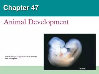
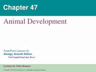
Chapter 47. Animal Development. 1 mm. Figure 47.1. A Body-Building Plan for Animals. Each of us began life as a single cell, a zygote A human embryo at approximately 6–8 weeks after conception Shows the development of distinctive features. Developmental stages. An organism’s development


Chapter 47
E N D
Presentation Transcript
Chapter 47 Animal Development
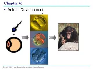
1 mm Figure 47.1 A Body-Building Plan for Animals • Each of us began life as a single cell, a zygote • A human embryo at approximately 6–8 weeks after conception • Shows the development of distinctive features
Developmental stages • An organism’s development • Is determined by the genome of the zygote and by differences that arise between early embryonic cells • After fertilization, embryonic development proceeds through cleavage, gastrulation, and organogenesis
Fertilization • The main function of fertilization • Is to bring the haploid nuclei of sperm and egg together to form a diploid zygote • Contact of the sperm with the egg’s surface • Initiates metabolic reactions within the egg that trigger the onset of embryonic development
Cleavage • Fertilization is followed by cleavage, a special type of cell division that creates the multicellular embryo from a single-celled zygote • During cleavage the cytoplasm pinches in, forming a cleavage furrow
Cleavage: Blastomeres and Morula stage • Cleavage partitions the cytoplasm of one large cell • into many smaller cells called blastomeres • the embryo does not actually grow in size ... morula - a solid ball of cells produced by continued cleavage
Cleavage: Blastula stage • blastula - hollow ball of cells formed when a fluid-filled cavity called the blastocoel forms within the morula
Animal and Vegetal Poles • The eggs and zygotes of many animals, except mammals • Have a definite polarity • The polarity is defined by the distribution of yolk • With the vegetal pole having the most yolk and the animal pole having the least • Cleavage planes usually follow a specific pattern • That is relative to the animal and vegetal poles of the zygote
Zygote 0.25 mm 2-cell stage forming Eight-cell stage (viewed from the animal pole). The large amount of yolk displaces the third cleavage toward the animal pole, forming two tiers of cells. The four cells near the animal pole (closer, in this view) are smaller than the other four cells (SEM). 4-cell stage forming 8-cell stage 0.25 mm Blasto- coel Animal pole Blastula (at least 128 cells). As cleavage continues, a fluid-filled cavity, the blastocoel, forms within the embryo. Because of unequal cell division due to the large amount of yolk in the vegetal hemisphere, the blastocoel is located in the animal hemisphere, as shown in the cross section. The SEM shows the outside of a blastula with about 4,000 cells, looking at the animal pole. Blastula (cross section) Vegetal pole Figure 47.9 Cleavage in a frog embryo
Gastrulation • The morphogenetic process called gastrulation • refers to the invagination or infolding of the blastula. • rearranges the cells of a blastula into a three-layered embryo, called a gastrula, that has a primitive gut Blastula Early Gastrula Late Gastrula
Embryonic Germ Layers • The three layers produced by gastrulation are called embryonic germ layers • The ectoderm forms the outermost layer of the gastrula • nervous system and epidermis • The endoderm forms the innermost layer of the gastrula • inner linings of the digestive tract and other systems • The mesoderm is the middle layer • muscle, skeleton, circulatory, reproductive, excretory, connective tissues
Organogenesis • Various regions of the three embryonic germ layers • Develop into the rudiments of organs during the process of organogenesis Eye Forebrain Heart Blood vessels Neural tube Figure 47.15b Late organogenesis. Rudiments of most major organs have already formed in this chick embryo, which is about 56 hours old and about 2–3 mm long (LM).
ECTODERM MESODERM ENDODERM • Epidermis of skin and itsderivatives (including sweatglands, hair follicles) • Epithelial lining of mouthand rectum • Sense receptors inepidermis • Cornea and lens of eye • Nervous system • Adrenal medulla • Tooth enamel • Epithelium or pineal andpituitary glands • Notochord • Skeletal system • Muscular system • Muscular layer of stomach, intestine, etc. • Excretory system • Circulatory and lymphaticsystems • Reproductive system(except germ cells) • Dermis of skin • Lining of body cavity • Adrenal cortex • Epithelial lining ofdigestive tract • Epithelial lining ofrespiratory system • Lining of urethra, urinarybladder, and reproductivesystem • Liver • Pancreas • Thymus • Thyroid and parathyroidglands Figure 47.16 Organogenesis and the 3 Germ Layers • Many different structures • Are derived from the three embryonic germ layers during organogenesis
Morphogenesis • Morphogenesis in animals involves specific changes in cell shape, position, and adhesion • Morphogenesis is a major aspect of development in both plants and animals, but only in animals does it involve the movement of cells • The cytoskeleton drives changes in the shape of the cell and cell migration, or cell crawling • Fibers of the extracellular matrix may function as tracks, directing migrating cells along particular routes
Ectoderm Neural plate Microtubules help elongate the cells of the neural plate. Microfilaments at the dorsal end of the cells may then contract,deforming the cells into wedge shapes. Cell wedging in the opposite direction causes the ectoderm to form a “hinge.” 1 4 2 3 Pinching off of the neural plate forms the neural tube. Figure 47.19 Change in cellular shape during morphogenesis
Cell Differentiation • Two general principles underlie differentiation during embryonic development • First, during early cleavage divisions • Embryonic cells must somehow become different from one another • Second, once initial cell asymmetries are set up • Interactions among the embryonic cells influence their fate, usually by causing changes in gene expression • This process is termed induction
Figure 21.11 Sources of developmental information for the early embryo (a) Cytoplasmic determinants in the egg. The unfertilized egg cell has molecules in its cytoplasm, encoded by the mother’s genes, that influence development. Many of these cytoplasmic determinants, like the two shown here, are unevenly distributed in the egg. After fertilization and mitotic division, the cell nuclei of the embryo are exposed to different sets of cytoplasmic determinants and, as a result, express different genes. Nucleus (b) Induction by nearby cells. The cells at the bottom of the early embryo depicted here are releasing chemicals that signal nearby cells to change their gene expression.
Restriction of Potency • In many species that have cytoplasmic determinants • Only the zygote is totipotent, capable of developing into all the cell types found in the adult