Chapter 47
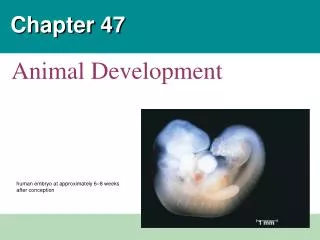
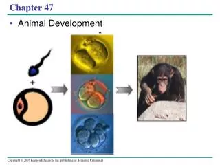
Chapter 47. Animal Development. LE 47-1. A human embryo at about 6–8. 1 mm. Important Steps in Development. Fertilization Cleavage Gastrulation Organogenesis. Acrosomal & Cortical Reactions In Sea Urchins. LE 47-3. Contact and fusion of sperm and egg membranes. Entry of sperm


Chapter 47
E N D
Presentation Transcript
Chapter 47 Animal Development
LE 47-1 A human embryo at about 6–8 1 mm
Important Steps in Development • Fertilization • Cleavage • Gastrulation • Organogenesis
Acrosomal & Cortical Reactions In Sea Urchins LE 47-3 Contact and fusion of sperm and egg membranes Entry of sperm nucleus Acrosomal reaction Sperm plasma membrane Sperm nucleus Cortical reaction Contact Acrosomal process Basal body (centriole) Sperm head Fertilization envelope Fused plasma membranes Cortical granule Actin Perivitelline space Hydrolytic enzymes Acrosome Jelly coat Cortical granule membrane Vitelline layer Sperm-binding receptors Egg plasma membrane EGG CYTOPLASM
LE 47-4 Cortical Reaction 500 µm 30 sec 10 sec after fertilization 20 sec 1 sec before fertilization Spreading wave of calcium ions Point of sperm entry
LE 47-6 Humans: Notice the Zona Pellucida Follicle cell Sperm basal body Cortical ganules Zona pellucida Sperm nucleus Egg plasma membrane Acrosomal vesicle EGG CYTOPLASM
LE 47-7 Cleavage • Fertilization is followed by cleavage, a period of rapid cell division without growth • Cleavage partitions the cytoplasm of one large cell into many smaller cells called blastomeres Fertilized egg Fertilized egg Four-cell stage Fertilized egg Morula Four-cell stage Four-cell stage Morula Blastula Morula Fertilized egg Blastula Four-cell stage Fertilized egg Four-cell stage Morula Blastula
LE 47-8 Animal hemisphere The eggs and zygotes of many animals, except mammals, have a definite polarity, defined by distribution of yolk. The development of body axes in frogs is influenced by the egg’s polarity Animal pole Point of sperm entry Vegetal hemisphere Vegetal pole Point of sperm entry Future dorsal side of tadpole Anterior Gray crescent Right First cleavage Ventral Dorsal Left Posterior Body axes Establishing the axes
LE 47-9 Zygote 0.25 mm 2-cell stage forming 4-cell stage forming Eight-cell stage (viewed from the animal pole) 8-cell stage 0.25 mm Animal pole Blasto- coel Blastula (cross section) Vegetal pole Blastula (at least 128 cells)
Gastrulation • Gastrulation rearranges the cells of a blastula into a three-layered embryo, called a gastrula, which has a primitive gut • The three layers produced by gastrulation are called embryonic germ layers • The ectoderm forms the outer layer • The endoderm lines the digestive tract • The mesoderm partly fills the space between the endoderm and ectoderm
LE 47-11 Key Gastrulation in a Sea Urchin Future ectoderm Future mesoderm Future endoderm Animal pole Blastocoel Mesenchyme cells Vegetal plate Vegetal pole Blastocoel Filopodia pulling archenteron tip Archenteron Mesenchyme cells Blastopore 50 µm Blastocoel Ectoderm Archenteron Blastopore Mouth Mesenchume (mesoderm forms future skeleton) Digestive tube (endoderm) Anus (from blastopore)
LE 47-12 CROSS SECTION SURFACE VIEW Gastrulation in a Frog Animal pole Blastocoel Dorsal tip of blastopore Dorsal lip of blastopore Vegetal pole Blastula Blastocoel shrinking Archenteron Ectoderm Mesoderm Blastocoel remnant Endoderm Key Future ectoderm Future mesoderm Yolk plug Yolk plug Gastrula Future endoderm
LE 47-14a Organogenesis Neural folds • During organogenesis, various regions of the germ layers develop into rudimentary organs • Early in vertebrate organogenesis, the notochord forms from mesoderm, and the neural plate forms from ectoderm LM 1 mm Neural fold Neural plate Notochord Ectoderm Mesoderm Endoderm Archenteron Neural plate formation
LE 47-14b Neural plate Neural fold • The neural plate soon curves inward, forming the neural tube Neural crest Outer layer of ectoderm Neural crest Neural tube Formation of the neural tube
LE 47-14c Somites Eye Tail bud SEM 1 mm Neural tube Notochord Neural crest Coelom Somite Archenteron (digestive cavity) Somites
Many structures are derived from the three embryonic germ layers during organogenesis
LE 47-17 Amnion Allantois Embryo Amniotic cavity with amniotic fluid Albumen Shell Yolk (nutrients) Yolk sac Chorion
Mammalian Development • Eggs of placental mammals show no obvious polarity • Early cleavage is relatively slow in humans and other mammals • At completion of cleavage, the blastocyst forms • The trophoblast, the outer epithelium of the blastocyst, initiates implantation in the uterus, and the blastocyst forms a flat disk of cells • As implantation is completed, gastrulation begins
LE 47-18a Endometrium (uterine lining) Inner cell mass Trophoblast Blastocoel Blastocyst reaches uterus. Expanding region of trophoblast Maternal blood vessel Epiblast Hypoblast Trophoblast Blastocyst implants.
Concept 47.3: The developmental fate of cells depends on their history and on inductive signals • Coupled with morphogenetic changes, development requires timely differentiation of cells at specific locations • Two general principles underlie differentiation: • During early cleavage divisions, embryonic cells must become different from one another • After cell asymmetries are set up, interactions among embryonic cells influence their fate, usually causing changes in gene expression
LE 47-23a Epidermis Central nervous system Epidermis Notochord Mesoderm Endoderm Neural tube stage (transverse section) Blastula Fate map of a frog embryo
LE 47-23b Cell lineage analysis in a tunicate
LE 47-26a Anterior The wings and legs of chicks, like all vertebrate limbs, begin as bumps of tissue called limb buds AER Limb bud ZPA Posterior Apical ectodermal ridge 50 µm Organizer regions
One limb-bud organizer region is the apical ectodermal ridge (AER) • The AER is thickened ectoderm at the bud’s tip • The second region is the zone of polarizing activity (ZPA) • The ZPA is mesodermal tissue under the ectoderm where the posterior side of the bud is attached to the body
LE 47-27 Anterior New ZPA Donor limb bud Host limb bud ZPA Posterior Tissue transplantation experiments support the hypothesis that the ZPA produces an inductive signal that conveys positional information indicating “posterior”