Download
1 / 114
1.18k likes | 1.51k Vues
Thromboelastometry - The New Coagulation Measurement . Andrew Bernard, MD Associate Professor of Surgery Trauma Center Medical Director UK Healthcare. Objectives. Understand how thromboelastometry works Describe the published literature on efficacy of TEM/TEG

E N D
Thromboelastometry- The New Coagulation Measurement Andrew Bernard, MD Associate Professor of Surgery Trauma Center Medical Director UK Healthcare
Objectives • Understand how thromboelastometry works • Describe the published literature on efficacy of TEM/TEG 3. Interpret typical thromboelastograms
Hypothesis: Pre-ICU MTP (FFP after 6 units PRBC) is inadequate for correcting coagulopathy.
Coagulopathy of Trauma Hemorrhagic State Bleeding Ongoing hypotension Pro-thrombotic State DVT / PE (Majority) Coagulopathy of trauma is dynamic.
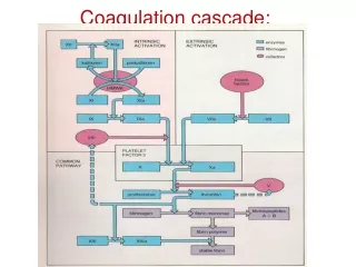
CONTACT TISSUE COMMON PATHWAY THROMBIN / FIBRINOGEN
Functional assay Global assessment (from initiation of protein coagulation through clot lysis) Factor Deficiencies Fibrinogen Function Platelet Function Clot Strength Lysis Thromboelastography
Thromboelastography Technology TEG ROTEM Delta
Thromboelastography Technology TEG ROTEM Delta
Hemostasis profile: R time Angle MA LY Fibrin strandsclot kineticsstrength/elasticitydissolution LY
R (reaction) time Coagulation factors K (clotting) time Interaction of factors, fibrin & platelets Alpha angle Fibrin & platelets Maximal Amplitude (MA) Platelet function Lysis 30/60 (LY30/60) Fibrinolysis TEG Components
R time • Sample placement to amplitude of 2mm • Clotting factor activity
K time • R time → amplitude of 20 mm • Time to reach level of clot strength • Clotting factors, fibrinogen, and platelet function
a-Angle • Slope from R-time to K-time • Speed of clot strengthening • Clotting factors, fibrinogen, and platelet function
Maximum Amplitude (MA) • Greatest vertical amplitude of tracing • Clot strength • Platelet number and function
LY30 • Rate of amplitude reduction @ 30 min after MA • Measure of clot stability • Increased rate indicates hyperfibrinolysis
GSW to Pelvis & RLE • Rectal, Small Bowel, Sacral, & Open Femur Fx • Arrived in Class IV Shock
Intra-op after 11 PRBC, 2 Plt, 4 Cryo, 6 FFP, 3 WB, & 1 Factor VIIa Post-op after 19 PRBC, 2 Plt, 4 Cryo, 6 FFP, 6 WB, & 1 Factor VIIa Same Patient
Patient 2 • L parietal GSW requiring emergent craniotomy • POD 1 : Hypercoagulable
GSW to Left Flank • Sigmoid Colon, Small Bowel, and Abdominal Wall Injury • 2 PRBC given intra-op
GSW to Left Flank • Post-op TEG shows early fibrinolysis • TEG after Amicar infusion
Clinical Randomisation of an Antifibrinolytic in Significant Haemorrhage
Guideline for Blood Product Use Abnormal TEG Prolonged R time Prolonged K time orDecrease a-Angle Transfuse 4 units FFP Transfuse 4 units FFP then 4 unitsCryoprecipitate Consider rVIIa if abnml after above Decrease Maximum Amplitude Increase LY30 Amicar 5gm IV load over 1 hourthen 1 gm/hr until LY normal Transfuse 2-4 units Whole Blood
Summary • Hemorrhage is the enemy (early) • Hypercoagulability is the enemy (late) • Diagnosis: time consuming and confusing • ROTEM Delta and TEG • “Whole blood coagulation measurement” • Fast • One test • Easily repeatable • It’s what you want-clot measurement
ROTEM delta • Proven technology - 1400 clinical units 50 countries -400 publications • Integrated system on-board computer, Touchscreen & LIS protocols • Electronic programmed Pipette & on screen step by step directions • LQC once a week, EQC automatic, CLIA Option 2 • Remote viewing in real time • Global Intrinsic & Extrinsic with specific Fibrinogen test
Surgical Hemostasis Webinar 4pm EDT Oct 20 Dr. Klaus Goerlinger
“Turnaround time” 2) “Obviation of other coagtests” 3) “Opportunity to return to POC (that we used to have with TEG in OR but had to abandon because of difficulty with instrumentation maintenance and QC)” 4) “Opportunity to use fewer units of clotting factors, thus improving outcome while saving money” 5) “As above, especially for platelet function (in future) so that not every patient on anti-platelet drugs has to get empiric platelets” 6) “Cost vsTEG”
Why United States last launch for Rotem? FDA Lawyers Rotem had to be Significantly better
Why Thromboelastometry ?Why Thromboelastometry?Target directed TransfusionsRapid reaction to coagulopathiesReduce Transfusion Costs…both Economic & Adverse Effects
ORIGINAL ARTICLE “An audit of red cell and blood product use after the institution of thromboelastometry in a cardiac intensive care unit” L. Anderson,* I. Quasim,* R. Soutar,† M. Steven,* A. Macfie* and W. Korte‡ *Department of Anaesthesia, †Department of Haematology, Western Infirmary, Glasgow, UK, and ‡Institute for Clinical Chemistry and Haematology, Kantonsspital, St Gallen, Switzerland Received 7 July 2005; accepted for publication 31 August 2005
A retrospective analysis of data from 990 patientswas performed which covered a period 6 months prior to the introduction of ROTEMthromboelastometry and 6 months after its introduction.
In the 6 months prior red cells were used in 60% of patients and fresh frozen plasma (FFP) and platelets used in 17 and 16% of patients, respectively. In the following 6 months red cell use had fallen to 53% and FFP and platelets to 12 and 11%, respectively (P < 005). Introduction of thromboelastometry has significantly decreased our use of red cells and blood products.
Annual treatment costs of all cardio surgical patients were analyzed before729 patients after 693 patients implementation of ‘bedside’ ROTEM.
Cost reduction of perioperative coagulation management in cardiac surgery: value of ‘bedside’ thrombelastography (ROTEM) Grit J. Spalding a, Martin Hartrumpf a, Tobias Sierig b, Nils Oesberg b, Christian Gu¨ntherKirschke b, Johannes M. Albes a,*Department of Cardiovascular Surgery, Heart Center Brandenburg, Bernau/Berlin, Germany Department of Anesthesiology, Heart Center Brandenburg, Bernau/Berlin, Germany Received 27 September 2006; received in revised form 26 February 2007; accepted 27 February 2007 Please cite this article in press as: Spalding GJ, et al., Cost reduction of perioperative coagulation management in cardiac surgery: value of ‘bedside’ thrombelastography (ROTEM), Eur J Cardio-thoracSurg (2007), doi:10.1016/j.ejcts.2007.02.022
After Rotem • Red Blood Cell Units < 25 % • Platelet Concentrates < 50% • FFP unchanged • Pooled coagulation concentrates & Factor XIII < 80% • Factor VIIA omitted • Fibrinogen > two fold • Avg. Monthly costs all blood products < 32% • Coagulation factor monthly costs 50% • Combined Savings 44%
ROTEM vs. TEG – Tests • TEG® • Koalin (intrinsic) • Heparinase cup • ROTEM® • IN-tem (intrinsic) • HEP-tem (heparinase) • Extem (extrinsic) • FIBtem (Fibrinogen ) • Aptem (Hyperfibrinolysis)